

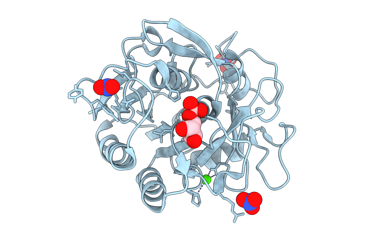
2ID8 image
Deposition Date 2006-09-14
Release Date 2006-10-10
Last Version Date 2024-10-30
Entry Detail
PDB ID:
2ID8
Keywords:
Title:
Crystal structure of Proteinase K
Biological Source:
Source Organism(s):
Method Details:
Experimental Method:
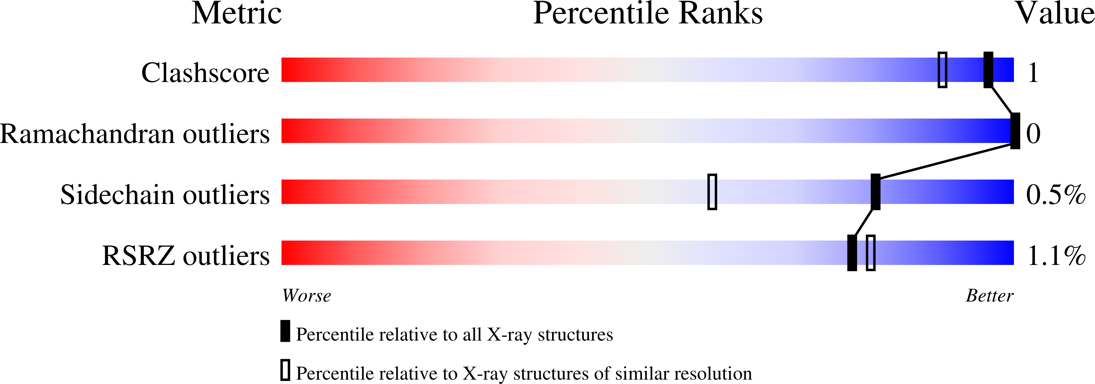
Resolution:
1.27 Å
R-Value Free:
0.15
R-Value Observed:
0.12
Space Group:
P 43 21 2
Macromolecular Entities
Polymer Type:polypeptide(L)
Molecule:Proteinase K
Gene (Uniprot):PROK
Chain IDs:A
Chain Length:279
Number of Molecules:1
Biological Source:Engyodontium album
Primary Citation
What can be done with a good crystal and an accurate beamline?
Acta Crystallogr. D Biol. Crystallogr. 62 1475 1483 (2006)
PMID: 17139083 DOI: 10.1107/S0907444906038534

Abstact

X-ray single-wavelength anomalous diffraction (SAD) data from a crystal of proteinase K were collected using synchrotron radiation of 0.98 A wavelength at SER-CAT 22-ID beamline, Advanced Photon Source, Argonne National Laboratory. At this wavelength, the expected Bijvoet ratio resulting from the presence of one calcium, one chloride and ten S atoms in the 279-residue protein is extremely small at approximately 0.46%. The direct-methods program SHELXD located 11 anomalous sites using data truncated to 2 A resolution. SHELXE was used to produce an easily interpretable electron-density map. This study shows that an accurate beamline and a good-quality crystal provide the possibility of successfully using a very weak anomalous signal of sulfur measured at a short wavelength for phasing a protein structure, even if a small degree of radiation damage is present.

Legend

Protein

Chemical

Disease

Primary Citation of related structures
Feedback Form
Name
Email
Institute
Feedback