

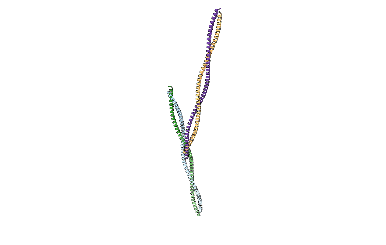
2EFR image
Deposition Date 2007-02-23
Release Date 2008-03-04
Last Version Date 2024-05-29
Entry Detail
PDB ID:
2EFR
Title:
Crystal structure of the c-terminal tropomyosin fragment with N- and C-terminal extensions of the leucine zipper at 1.8 angstroms resolution
Biological Source:
Source Organism(s):
Expression System(s):
Method Details:
Experimental Method:
Resolution:
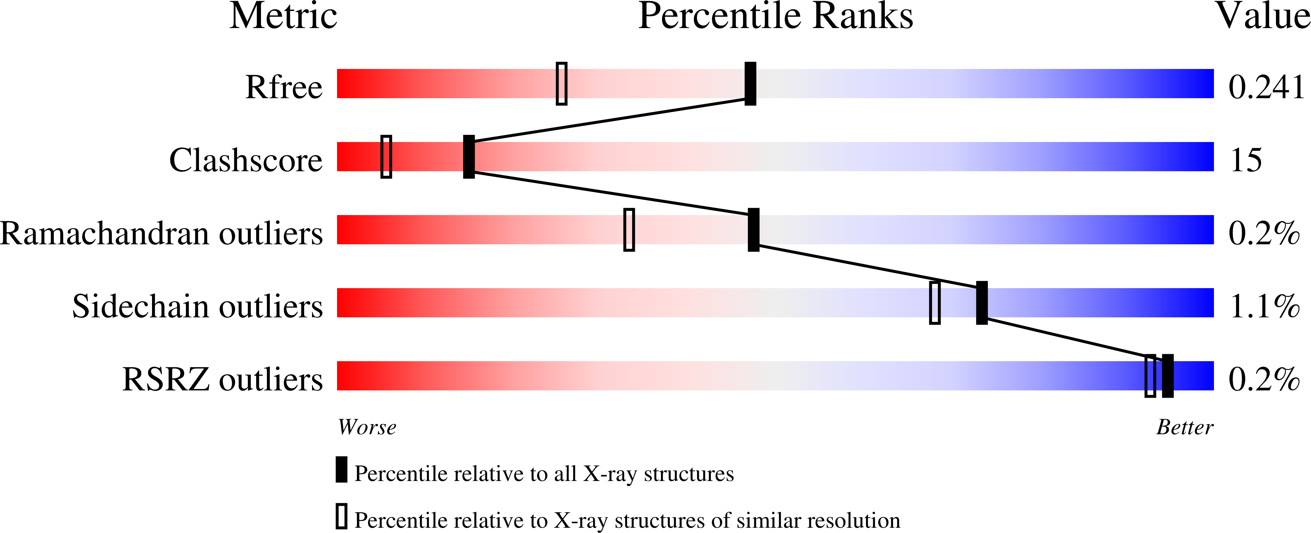
1.80 Å
R-Value Free:
0.31
R-Value Work:
0.23
R-Value Observed:
0.23
Space Group:
P 1
Macromolecular Entities
Protein Blast
Polymer Type:polypeptide(L)
Molecule:General control protein GCN4
Gene (Uniprot):GCN4, TPM1
Mutagens:C190S
Chain IDs:A, B, C, D
Chain Length:155
Number of Molecules:4
Biological Source:Saccharomyces cerevisiae, Oryctolagus cuniculus
Primary Citation
Two-crystal structures of tropomyosin C-terminal fragment 176-273: exposure of the hydrophobic core to the solvent destabilizes the tropomyosin molecule.
Biophys J 95 710 ? (2008)
PMID: 18339732 DOI: .

Abstact

Tropomyosin (Tm) is a two-stranded alpha-helical coiled-coil protein, and when associated with troponin, it is responsible for the actin filament-based regulation of muscle contraction in vertebrate skeletal and cardiac muscles. It is widely believed that Tm adopts a flexible rod-like structure in which the flexibility must play a crucial role in its functions. To obtain more information about the flexibility of Tm, we solved and compared two crystal structures of the identical C-terminal segments, spanning approximately 40% of the entire length. We also compared these structures with our previously reported crystal structure of an almost identical Tm segment in a distinct crystal form. The parameters specifying the local coiled-coil geometry, such as the separation between two helices and the local helical pitch, undulate along the length of Tm in the same way as among the three crystal structures, indicating that these parameters are defined by the amino acid sequence. In the region of increased separation, around Glu-218 and Gln-263, the hydrophobic core is disrupted by three holes. Moreover, for the first time to our knowledge, for Tm, water molecules have been identified in these holes. In some structures, the B-factors are higher around the holes than in the rest of the molecule. The Tm coiled-coil must be destabilized and therefore may be flexible, not only in the alanine clusters but also in the regions of the broken core. A closer look at the local staggering between the two chains and the local bending revealed that the strain accumulates at the alanine cluster and may be relaxed in the broken core region. Moreover, the strain is distributed over a long range, even when a deformation like bending may occur at a limited number of spots. Thus, Tm should not be regarded as a train of short rigid rods connected by flexible linkers, but rather as a seamless rubber rod patched with relatively more flexible regions.

Legend

Protein

Chemical

Disease

Primary Citation of related structures
Feedback Form
Name
Email
Institute
Feedback