2BD0 image
Deposition Date 2005-10-19
Release Date 2005-11-29
Last Version Date 2024-03-13
Entry Detail
PDB ID:
2BD0
Keywords:
Title:
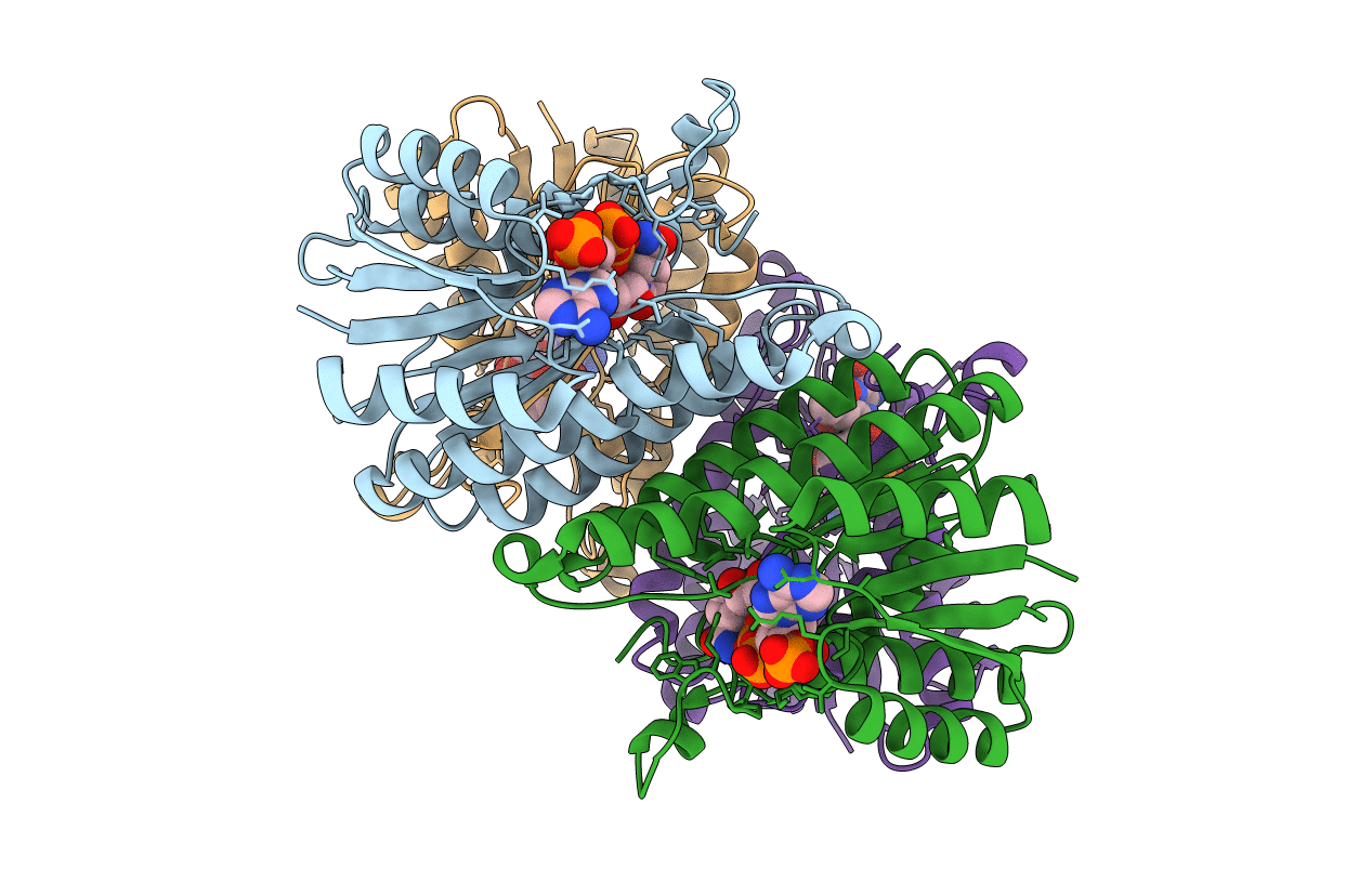
Chlorobium tepidum Sepiapterin Reductase complexed with NADP and Sepiapterin
Biological Source:
Source Organism(s):
Expression System(s):
Method Details:
Experimental Method:
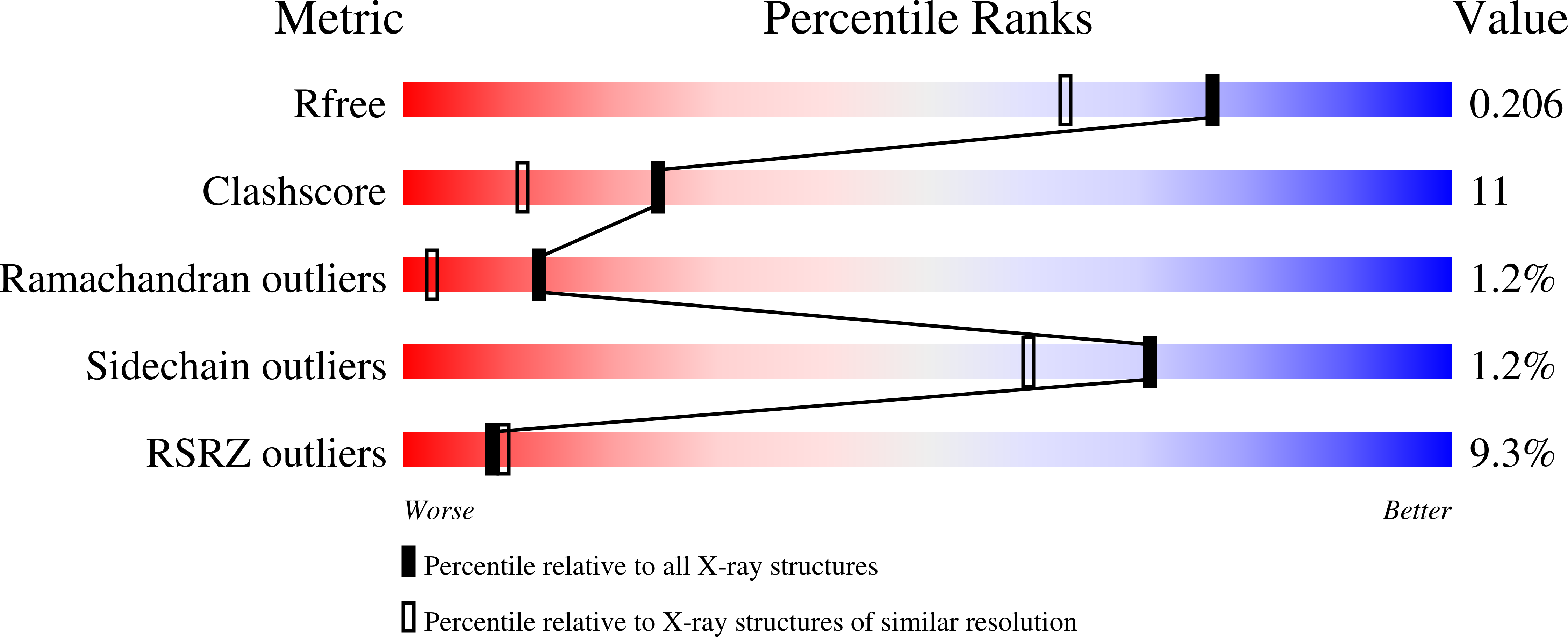
Resolution:
1.70 Å
R-Value Free:
0.21
R-Value Work:
0.2
Space Group:
P 21 21 21
Macromolecular Entities
Structures with similar UniProt ID
Protein Blast
Polymer Type:polypeptide(L)
Molecule:sepiapterin reductase
Gene (Uniprot):CT0609
Chain IDs:A, B, C, D
Chain Length:244
Number of Molecules:4
Biological Source:Chlorobium tepidum
Primary Citation
Structure of Chlorobium tepidum sepiapterin reductase complex reveals the novel substrate binding mode for stereospecific production of L-threo-tetrahydrobiopterin
J. Biol. Chem. 281 2249 2256 (2006)
PMID: 16308317 DOI: 10.1074/jbc.M509343200

Abstact

Sepiapterin reductase (SR) is involved in the last step of tetrahydrobiopterin (BH(4)) biosynthesis by reducing the di-keto group of 6-pyruvoyl tetrahydropterin. Chlorobium tepidum SR (cSR) generates a distinct BH(4) product, L-threo-BH(4) (6R-(1'S,2'S)-5,6,7,8-BH(4)), whereas animal enzymes produce L-erythro-BH(4) (6R-(1'R,2'S)-5,6,7,8-BH(4)) although it has high amino acid sequence similarities to the other animal enzymes. To elucidate the structural basis for the different reaction stereospecificities, we have determined the three-dimensional structures of cSR alone and complexed with NADP and sepiapterin at 2.1 and 1.7 A resolution, respectively. The overall folding of the cSR, the binding site for the cofactor NADP(H), and the positions of active site residues were quite similar to the mouse and the human SR. However, significant differences were found in the substrate binding region of the cSR. In comparison to the mouse SR complex, the sepiapterin in the cSR is rotated about 180 degrees around the active site and bound between two aromatic side chains of Trp-196 and Phe-99 so that its pterin ring is shifted to the opposite side, but its side chain position is not changed. The swiveled sepiapterin binding results in the conversion of the side chain configuration, exposing the opposite face for hydride transfer from NADPH. The different sepiapterin binding mode within the conserved catalytic architecture presents a novel strategy of switching the reaction stereospecificities in the same protein fold.

Legend

Protein

Chemical

Disease

Primary Citation of related structures
Feedback Form
Name
Email
Institute
Feedback