

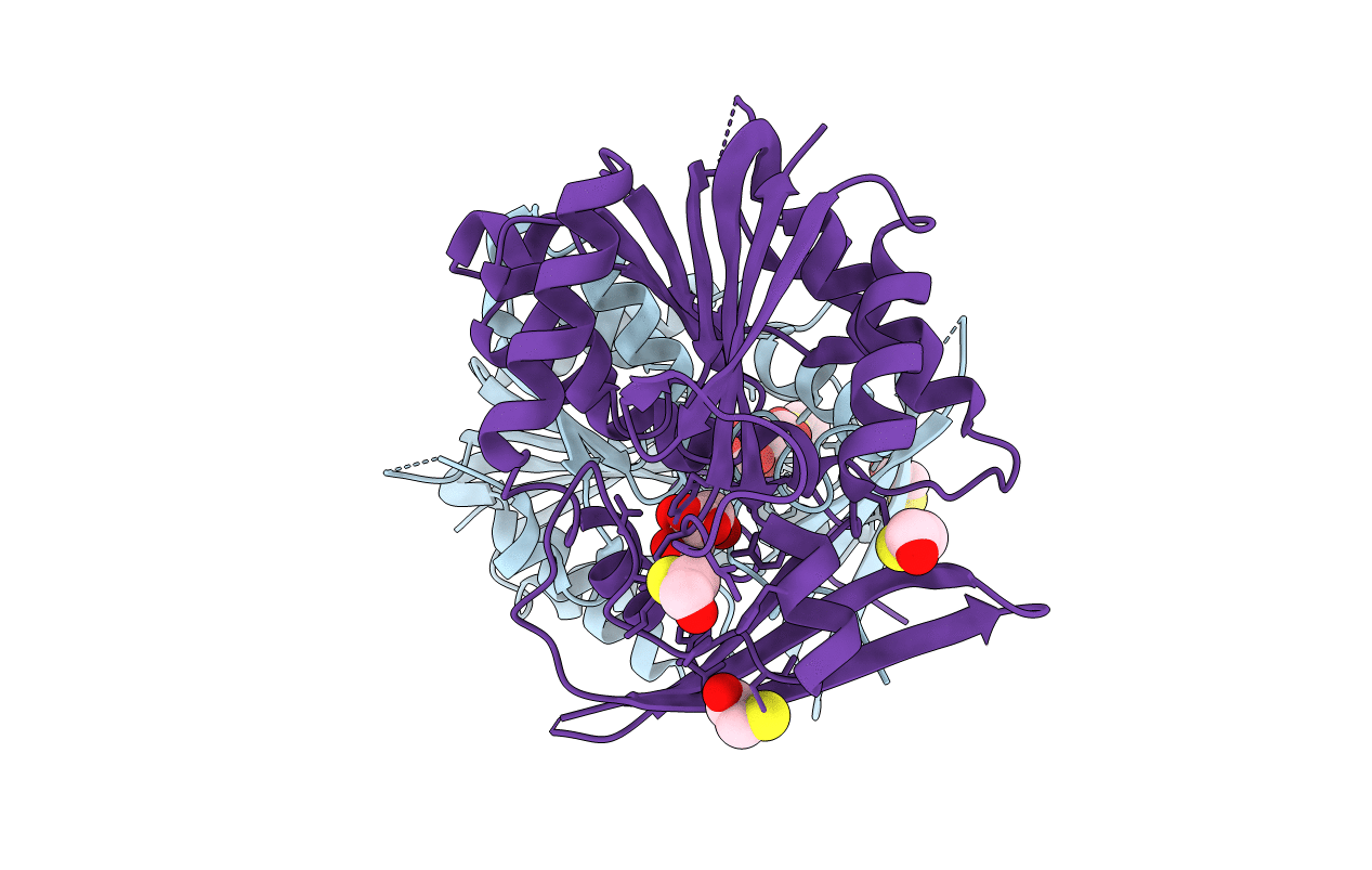
2AZT image
Deposition Date 2005-09-12
Release Date 2006-09-26
Last Version Date 2023-08-23
Entry Detail
PDB ID:
2AZT
Keywords:
Title:
Crystal structure of H176N mutant of human Glycine N-Methyltransferase
Biological Source:
Source Organism(s):
Homo sapiens (Taxon ID: 9606)
Expression System(s):
Method Details:
Experimental Method:
Resolution:
2.70 Å
R-Value Free:
0.28
R-Value Work:
0.23
R-Value Observed:
0.23
Space Group:
P 21 21 2

Macromolecular Entities
Structures with similar UniProt ID
Protein Blast
Polymer Type:polypeptide(L)
Molecule:Glycine N-methyltransferase
Gene (Uniprot):GNMT
Mutagens:H176N
Chain IDs:A, B
Chain Length:295
Number of Molecules:2
Biological Source:Homo sapiens
Primary Citation
Destabilization of human glycine N-methyltransferase by H176N mutation.
Protein Sci. 16 1957 1964 (2007)
PMID: 17660255 DOI: 10.1110/ps.072921507

Abstact

In the presence of moderate (2-4 M) urea concentrations the tetrameric enzyme, glycine N-methyltransferase (GNMT), dissociates into compact monomers. Higher concentrations of urea (7-8 M) promote complete denaturation of the enzyme. We report here that the H176N mutation in this enzyme, found in humans with hypermethioninaemia, significantly decreases stability of the tetramer, although H176 is located far from the intersubunit contact areas. Dissociation of the tetramer to compact monomers and unfolding of compact monomers of the mutant protein were detected by circular dichroism, quenching of fluorescence emission, size-exclusion chromatography, and enzyme activity. The values of apparent free energy of dissociation of tetramer and of unfolding of compact monomers for the H176N mutant (27.7 and 4.2 kcal/mol, respectively) are lower than those of wild-type protein (37.5 and 6.2 kcal/mol). A 2.7 A resolution structure of the mutant protein revealed no significant difference in the conformation of the protein near the mutated residue.

Legend

Protein

Chemical

Disease

Primary Citation of related structures
Feedback Form
Name
Email
Institute
Feedback