

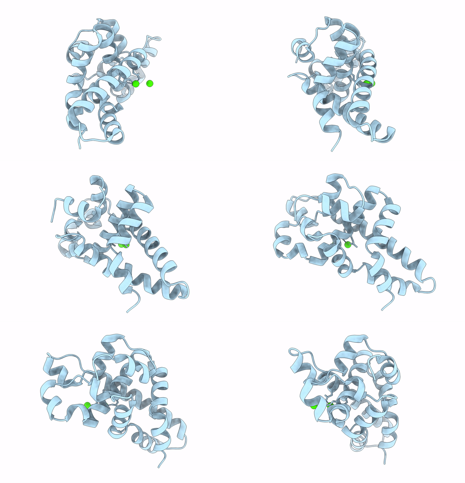
2A11 image
Deposition Date 2005-06-17
Release Date 2005-07-05
Last Version Date 2024-02-14
Entry Detail
PDB ID:
2A11
Title:
Crystal Structure of Nuclease Domain of Ribonuclase III from Mycobacterium Tuberculosis
Biological Source:
Source Organism(s):
Expression System(s):
Method Details:
Experimental Method:
Resolution:
2.10 Å
R-Value Free:
0.28
R-Value Work:
0.25
R-Value Observed:
0.28
Space Group:
P 43 21 2

Macromolecular Entities
Polymer Type:polypeptide(L)
Molecule:Ribonuclease III
Chain IDs:A
Chain Length:242
Number of Molecules:1
Biological Source:Mycobacterium tuberculosis
Ligand Molecules
Primary Citation
Structure of the nuclease domain of ribonuclease III from M. tuberculosis at 2.1 A
Protein Sci. 14 2744 2750 (2005)
PMID: 16155207 DOI: 10.1110/ps.051665905

Abstact

RNase III enzymes are a highly conserved family of proteins that specifically cleave double-stranded (ds)RNA. These proteins are involved in a diverse group of functions, including ribosomal RNA processing, mRNA maturation and decay, snRNA and snoRNA processing, and RNA interference. Here we report the crystal structure of the nuclease domain of RNase III from the pathogen Mycobacterium tuberculosis. Although globally similar to other RNase III folds, this structure has some features not observed in previously reported models. These include the presence of an additional metal ion near the catalytic site, as well as conserved secondary structural elements that are proposed to have functional roles in the recognition of dsRNAs.

Legend

Protein

Chemical

Disease

Primary Citation of related structures
Feedback Form
Name
Email
Institute
Feedback