

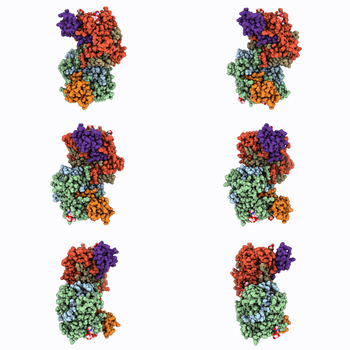
29WJ image
Deposition Date 2026-04-10
Release Date 2026-10-07
Last Version Date 2026-10-14
Entry Detail
PDB ID:
29WJ
Keywords:
Title:
Structure of monomerized native leukocyte myeloperoxidase in complex with the Staphylococcal Peroxidase Inhibitor SPIN
Biological Source:
Source Organism(s):
Expression System(s):
Method Details:
Experimental Method:
Resolution:
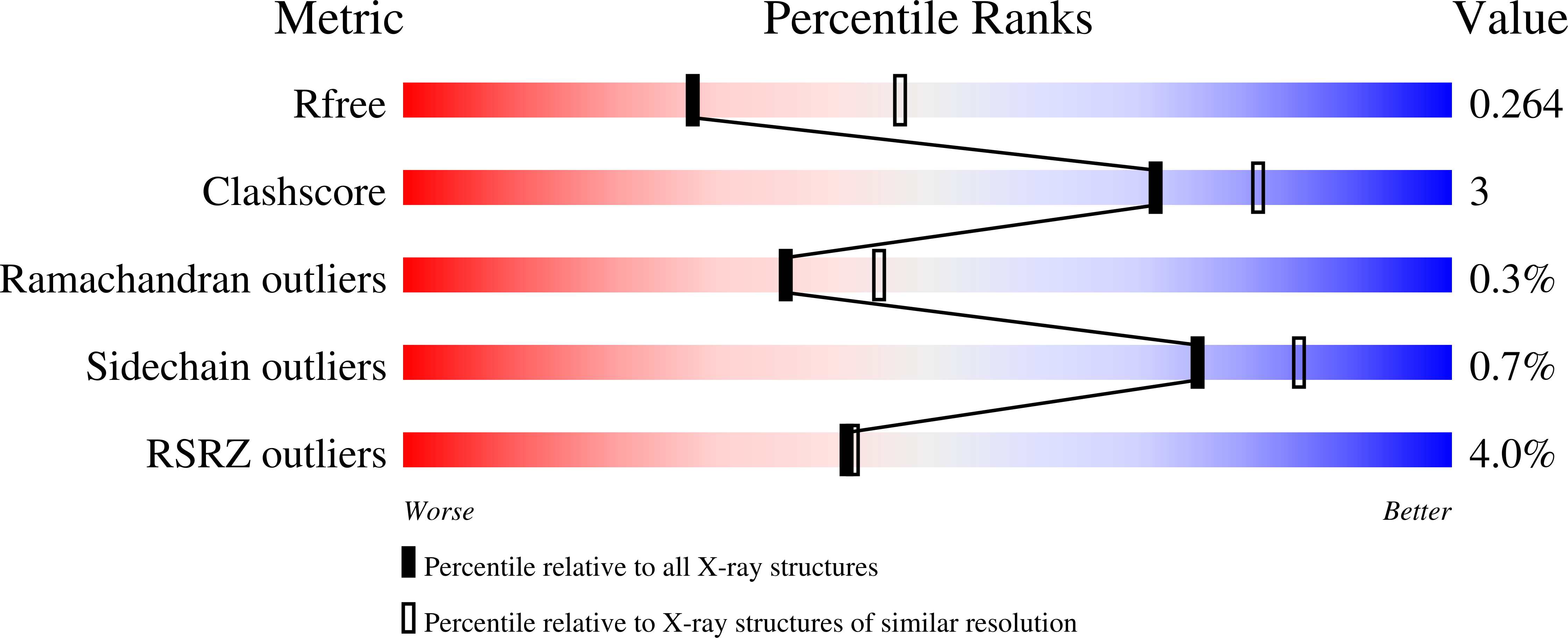
2.55 Å
R-Value Free:
0.26
R-Value Work:
0.20
R-Value Observed:
0.20
Space Group:
P 43 21 2
Macromolecular Entities
Polymer Type:polypeptide(L)
Molecule:Myeloperoxidase
Gene (Uniprot):MPO
Chain IDs:A, D (auth: C)
Chain Length:114
Number of Molecules:2
Biological Source:Homo sapiens
Polymer Type:polypeptide(L)
Molecule:Myeloperoxidase
Gene (Uniprot):MPO
Chain IDs:B
Chain Length:467
Number of Molecules:1
Biological Source:Homo sapiens
Polymer Type:polypeptide(L)
Molecule:Myeloperoxidase
Gene (Uniprot):MPO
Chain IDs:E (auth: D)
Chain Length:467
Number of Molecules:1
Biological Source:Homo sapiens
Polymer Type:polypeptide(L)
Molecule:Myeloperoxidase inhibitor SPI
Gene (Uniprot):SAOUHSC_00401
Chain IDs:C (auth: E), F
Chain Length:73
Number of Molecules:2
Biological Source:Staphylococcus aureus
Modified Residue
Compound ID Chain ID Parent Comp ID Details 2D Image
YCM A, B, D, E CYS modified residue
Primary Citation
Interface N-glycans drive myeloperoxidase dimerization in vitro.
Int.J.Biol.Macromol. 383 154499 154499 (2026)
PMID: 42735772 DOI: 10.1016/j.ijbiomac.2026.154499

Abstact

Myeloperoxidase (MPO) is a highly glycosylated heme oxidoreductase that, together with lactoperoxidase (LPO) and eosinophil peroxidase (EPO), contributes to host defence through the generation of (pseudo)hypohalous acids. Among the human heme peroxidases, mature MPO forms a unique covalently linked homodimer. Dimerization enhances stability and is essential for some biological functions, including chromatin disruption during neutrophil extracellular trap formation. Thus, we hypothesized that MPO dimerization must reflect a tightly regulated step in MPO biosynthesis and is driven by specific structural elements. Cross-species sequence alignment indicated that N-glycosylation motifs were evolutionarily more conserved in MPO than in monomeric EPO and LPO. We investigated the role of N-glycans in MPO dimerization using in vitro monomerized glycosylated (mMPO) and deglycosylated (mMPOdg) monomeric variants of native human MPO. Small-angle X-ray scattering, isothermal titration calorimetry and crystal structures showed that glycosylated MPO monomers had a weak but biologically relevant affinity in the millimolar range, which was lost upon deglycosylation. Furthermore, we present the first cryogenic transmission electron microscopy structure of human MPO with at least one core N-acetylglucosamine residue resolved at each N-glycosylation site. Structural analysis suggests that an extensive hydrogen bonding network of interface N-glycans at position 483 and residues of the other monomer drives MPO homodimerization. Dimerization enhances stability without affecting enzymatic activity. Together, we show that N-glycans are essential to MPO dimerization, provide important insights into the impact of interface N-glycans on biophysical and biochemical properties of MPO and discuss the implications for the biological roles of MPO in health and disease.

Legend

Protein

Chemical

Disease

Primary Citation of related structures
Feedback Form
Name
Email
Institute
Feedback