28PB image
Deposition Date 2026-02-12
Release Date 2026-05-27
Last Version Date 2026-06-10
Entry Detail
PDB ID:
28PB
Title:
Crystal structure of CbcA periplasmic domain from Geobacter sulfurreducens
Biological Source:
Source Organism(s):
Expression System(s):
Method Details:
Experimental Method:
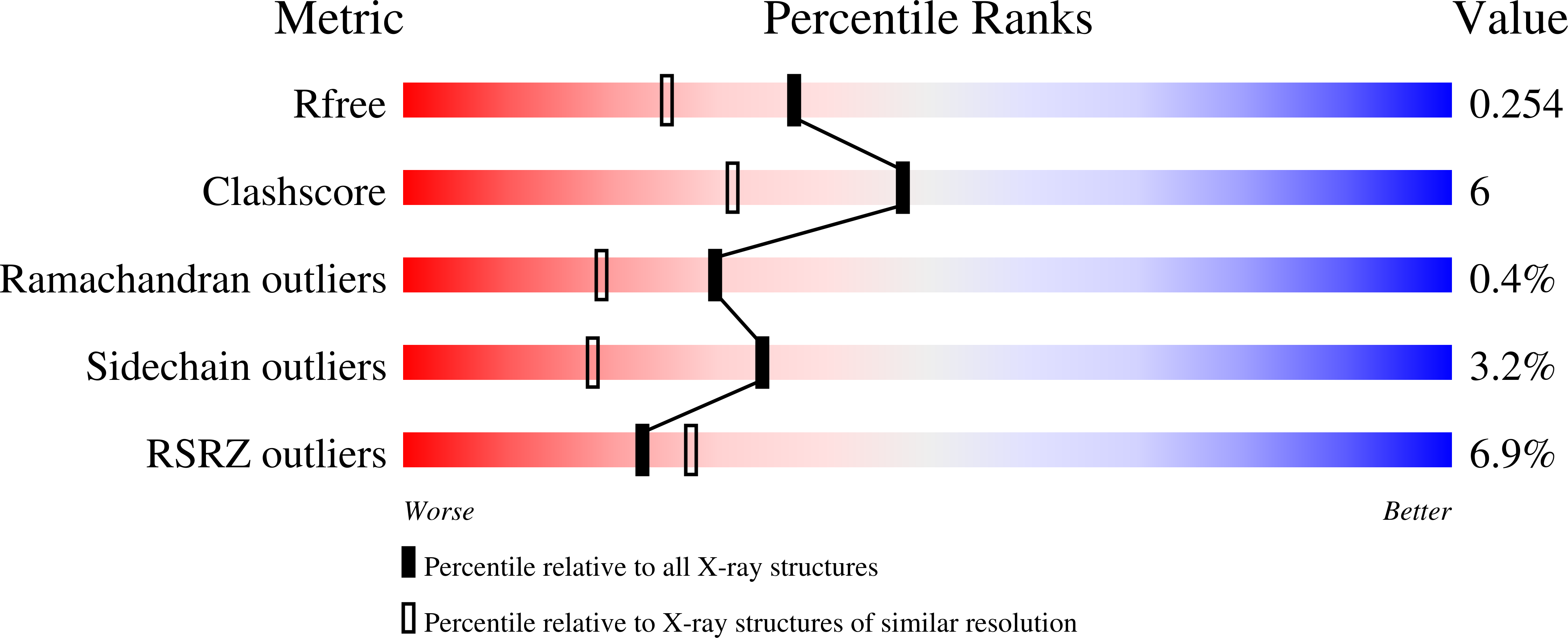
Resolution:
1.91 Å
R-Value Free:
0.25
R-Value Work:
0.19
Space Group:
P 21 21 21
Macromolecular Entities
Structures with similar UniProt ID
Protein Blast
Polymer Type:polypeptide(L)
Molecule:Cytochrome c
Gene (Uniprot):GSU0594
Chain IDs:A, B
Chain Length:278
Number of Molecules:2
Biological Source:Geobacter sulfurreducens PCA
Primary Citation
Bridging inner membrane and periplasm in Geobacter sulfurreducens: structural and biochemical insights into CbcA and its redox partners.
Int.J.Biol.Macromol. 369 152597 152597 (2026)
PMID: 42155738 DOI: 10.1016/j.ijbiomac.2026.152597

Abstact

Biotechnological applications such as bioremediation, bioenergy production, and microbial electrosynthesis are emerging as sustainable alternatives to conventional and environmentally harmful industrial practices. Advancing these technologies requires a deeper understanding of extracellular electron transfer, a process mediated by a network of redox partners bridging the inner membrane and the extracellular environment in electroactive bacteria. The CbcBA complex, a quinol:cytochrome c oxidoreductase from Geobacter sulfurreducens, is essential for the reduction of extracellular metal oxides and electrodes with redox potential values below -210 mV. The complex comprises CbcA, a heptaheme c-type cytochrome anchored to the inner membrane and CbcB, an integral membrane di-heme b-type cytochrome. Additionally, CbcC, a periplasmic dodecaheme cytochrome, and the five periplasmic triheme cytochromes PpcA-E, are proposed to function as redox partners of CbcBA. To investigate their structural and functional properties, the periplasmic domain of CbcA (CbcA(sol)) and CbcC were heterologously expressed and analyzed using complementary spectroscopic techniques. The crystal structure of CbcA(sol) was solved at 1.9 A resolution, revealing a calcium-binding EF-hand motif that may function as a regulatory switch. Circular dichroism and differential scanning calorimetry indicated that CbcA(sol) and CbcC exhibit high stability, while potentiometric redox titrations demonstrated distinct electrochemical behaviors: CbcA(sol) has the most negative reduction potential among G. sulfurreducens oxidoreductases, whereas CbcC operates within the redox range of the PpcA-E cytochromes. NMR experiments showed that CbcA(sol) transfers electrons to CbcC and PpcA-E cytochromes. This result, in agreement with the structural electrostatic complementarity between the different cytochromes, suggests that CbcA may interact with multiple periplasmic cytochromes through distinct surface regions.

Legend

Protein

Chemical

Disease

Primary Citation of related structures
Feedback Form
Name
Email
Institute
Feedback