

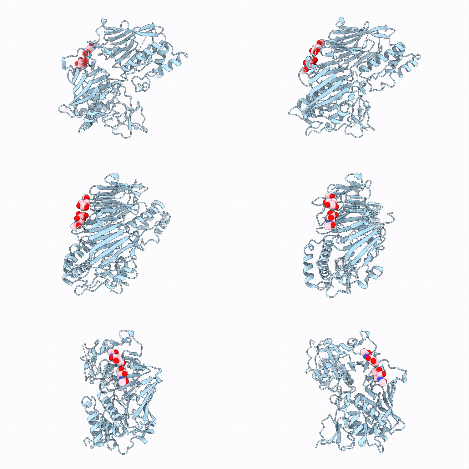
24NE image
Deposition Date 2026-03-12
Release Date 2026-04-08
Last Version Date 2026-04-08
Entry Detail
PDB ID:
24NE
Title:
Crystal Structure of Cypridina luciferase
Biological Source:
Source Organism(s):
Expression System(s):
Method Details:
Experimental Method:
Resolution:
3.00 Å
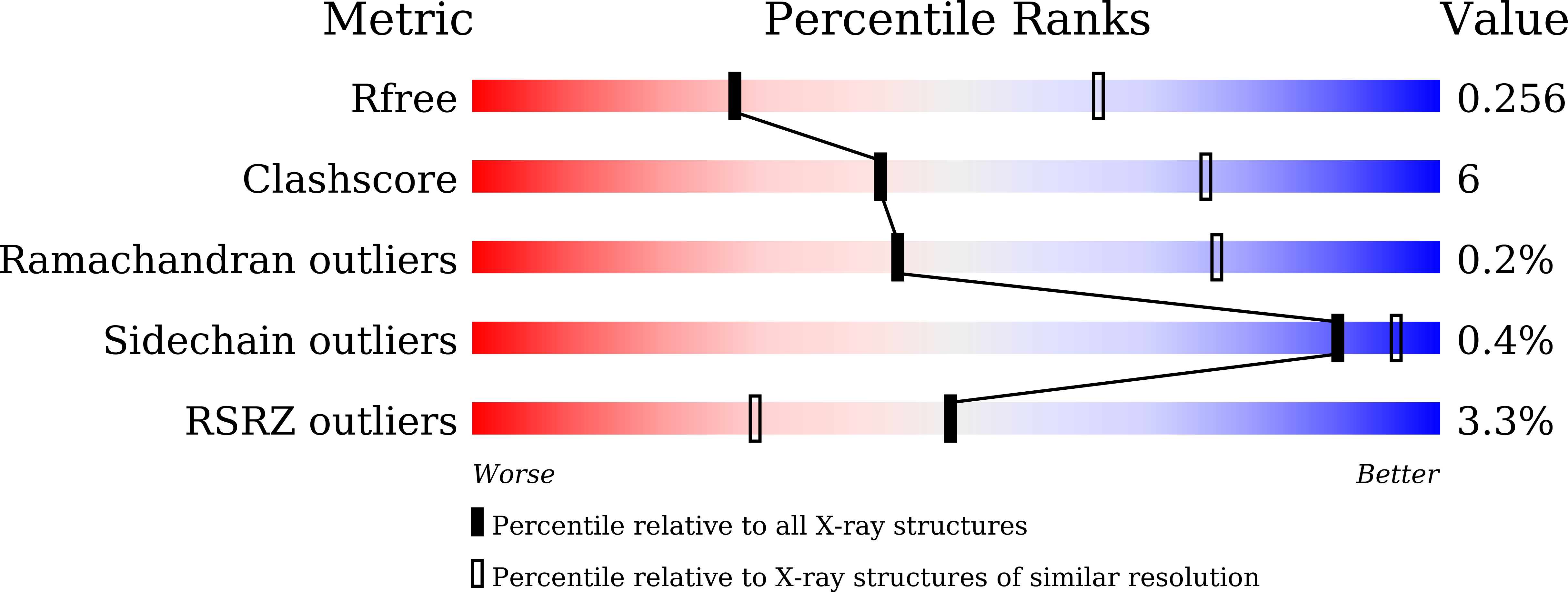
R-Value Free:
0.26
R-Value Work:
0.23
R-Value Observed:
0.23
Space Group:
P 63 2 2
Macromolecular Entities
Structures with similar UniProt ID
Protein Blast
Polymer Type:polypeptide(L)
Molecule:Luciferase
Gene (Uniprot):CnL
Chain IDs:A
Chain Length:537
Number of Molecules:1
Biological Source:Cypridina noctiluca
Ligand Molecules
Primary Citation
Three-dimensional structure of Cypridina luciferase illuminates the mechanism of bioluminescence of imidazopyrazinone-type luciferin.
Int. J. Biol. Macromol. 357 151583 151583 (2026)
PMID: 41865923 DOI: 10.1016/j.ijbiomac.2026.151583

Abstact

Bioluminescence occurs in marine organisms and involves the oxidation of imidazopyrazinone-type luciferin catalyzed by luciferase. Although chemiluminescence mechanisms have been studied using luciferin analogs in aprotic solvents, the enzymatic reaction within luciferase remains poorly understood due to the lack of structural information. Cypridina luciferin (CypL), used by Vargula hilgendorfii, is an imidazopyrazinone compound known for its high quantum yield and turnover rate. Here, we report the crystal structure of Cypridina luciferase (CLuc) and its reaction product, Cypridina oxyluciferin (CypOL). Structural analysis and mutagenesis suggest that the amino acid residue H542 may mediate either deprotonation at the N7 position or stabilization of the anionic form of CypL in the first step of the oxidation process. This mechanism, together with strict substrate recognition, would provide a clue to investigate the molecular basis of CLuc's efficient light emission and advance understanding of imidazopyrazinone-type bioluminescence.

Legend

Protein

Chemical

Disease

Primary Citation of related structures
Feedback Form
Name
Email
Institute
Feedback