

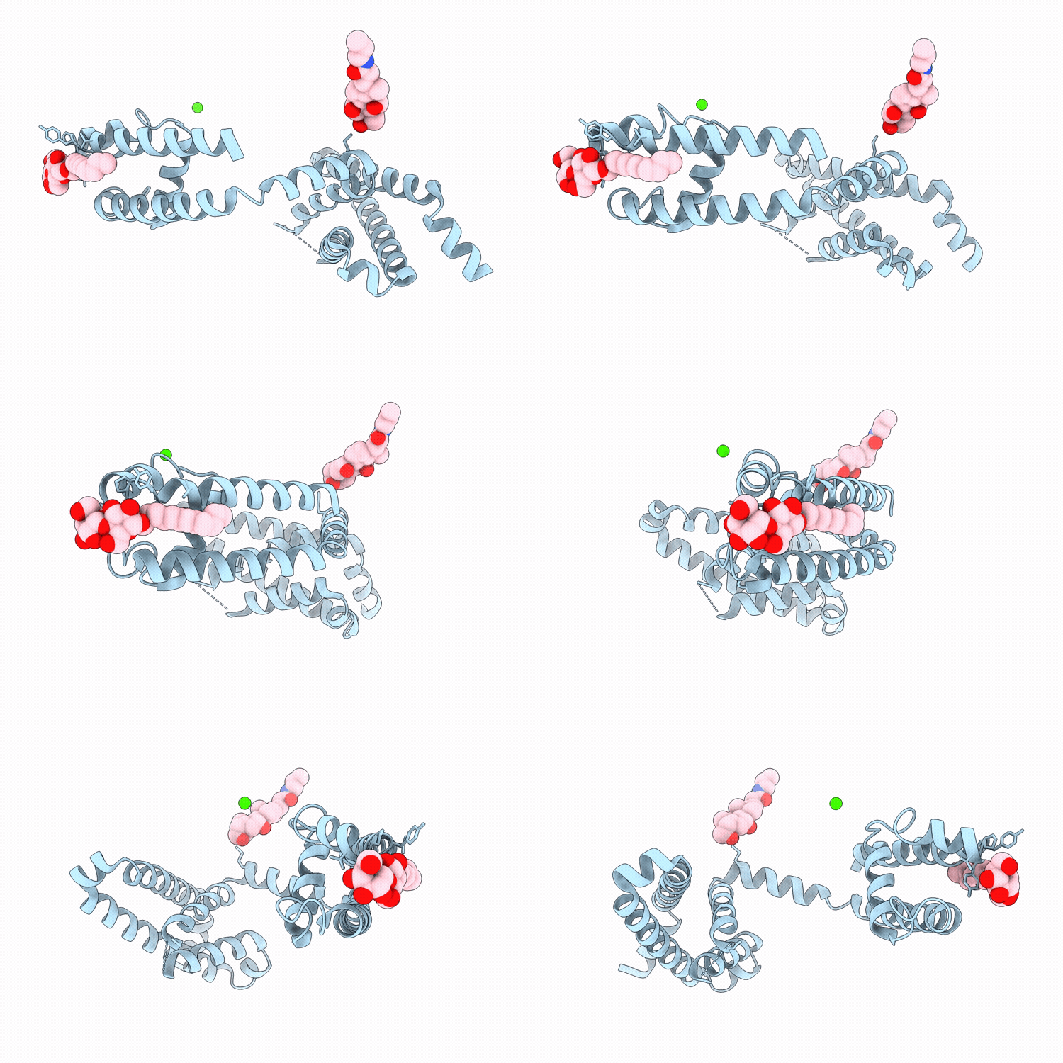
24JT image
Deposition Date 2026-03-06
Release Date 2026-05-20
Last Version Date 2026-05-27
Entry Detail
PDB ID:
24JT
Title:
Crystal structure of voltage-gated sodium channel NavAb N49K mutant
Biological Source:
Source Organism(s):
Expression System(s):
Method Details:
Experimental Method:
Resolution:
4.10 Å
R-Value Free:
0.37
R-Value Work:
0.35
R-Value Observed:
0.35
Space Group:
I 4 2 2
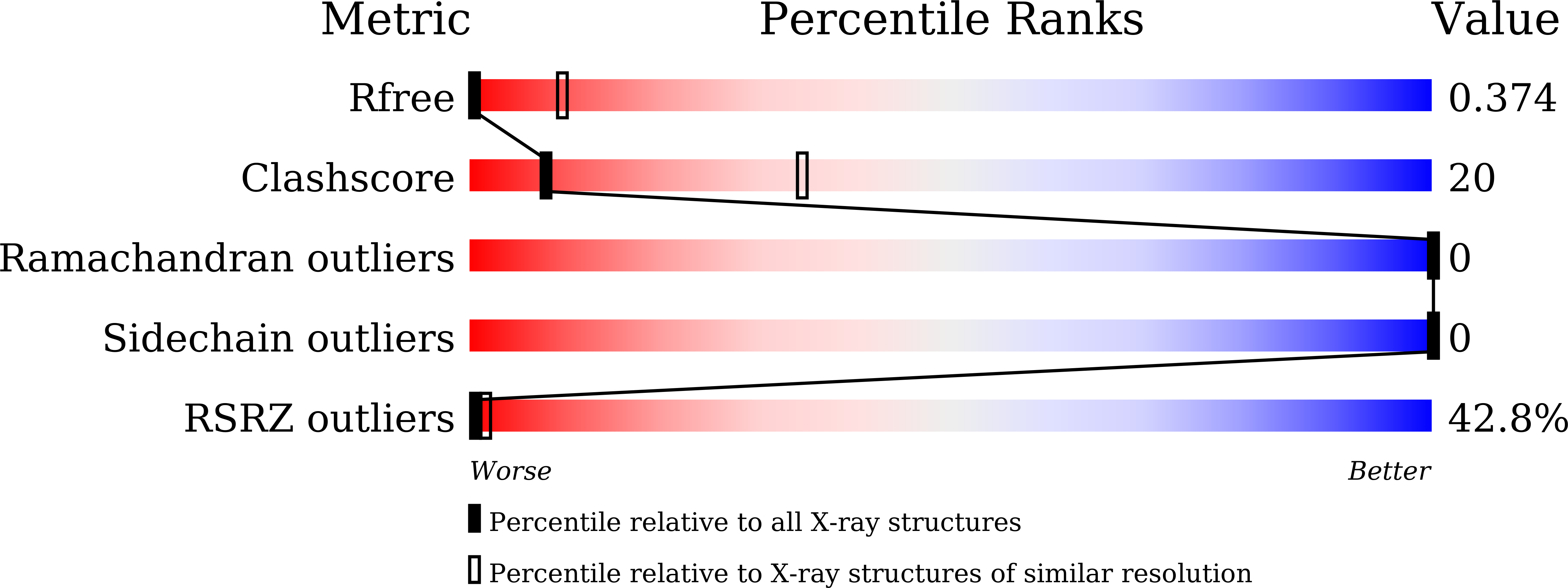
Macromolecular Entities
Structures with similar UniProt ID
Protein Blast
Polymer Type:polypeptide(L)
Molecule:Ion transport protein
Gene (Uniprot):Abu_1752
Mutagens:N49K
Chain IDs:A
Chain Length:229
Number of Molecules:1
Biological Source:Aliarcobacter butzleri RM4018
Primary Citation
Conformational dynamics underlying slow inactivation in voltage-gated sodium channels.
Biorxiv ? ? ? (2025)
PMID: 40894622 DOI: 10.1101/2025.08.14.670348

Abstact

Voltage-gated sodium (Nav) channels initiate and propagate action potentials in many excitable cells. Upon repetitive activation, the fraction of Nav channels available for excitation gradually decreases on a timescale ranging from seconds to minutes, a phenomenon known as slow inactivation. This process is crucial for regulating cellular excitability and firing patterns. Slow inactivation is proposed to result from the collapse of the selectivity filter pore coupled with the opening of the primary helix bundle crossing gate. However, the conformational changes underlying slow inactivation and the molecular coupling between the selectivity filter and primary gate remain unclear. In this study, we investigated the conformational dynamics of the selectivity filter in prokaryotic NavAb channels reconstituted into liposomes using single-molecule FRET (smFRET). Our smFRET data revealed the conformational transitions in the NavAb selectivity filter pore among three distinct states, with activating voltages enriching the high-FRET conformations, potentially associated with slow inactivation. Using electrophysiological and crystallographic methods, we further identified the L176 residue in the selectivity filter P1 helix as a critical coupler between the primary and slow inactivation gates. We showed that L176 mutations with side chains of larger sizes significantly facilitated the slow inactivation of the NavAb channel, and the L176F mutation forced the opening mutant carrying the C-terminal deletion to be crystallized at the closed state. Consistently, our smFRET results revealed that C-terminal deletion markedly attenuated the high FRET conformation of the selectivity filter, which was restored by the L176F mutation. Moreover, using the classical Nav open-pore blocker lidocaine, we showed that it also depleted the high FRET conformation of the NavAb selectivity filter in a dose-dependent manner. The L176F mutation, again, markedly reversed the conformational shifts caused by lidocaine, an effect similar to it on the opening mutant carrying the C-terminal deletion. Our studies consistently suggested that slow inactivation in the NavAb channel is underlined by the collapse of the selectivity filter pore, represented by the high FRET conformation uncovered by our smFRET measurements, while the L176 residue at the P1 helix of the selectivity filter and T206 at the pore lining helix couple the conformational changes of the slow inactivation gate at selectivity filter and the primary gate at the helix bundle crossing.

Legend

Protein

Chemical

Disease

Primary Citation of related structures
Feedback Form
Name
Email
Institute
Feedback