

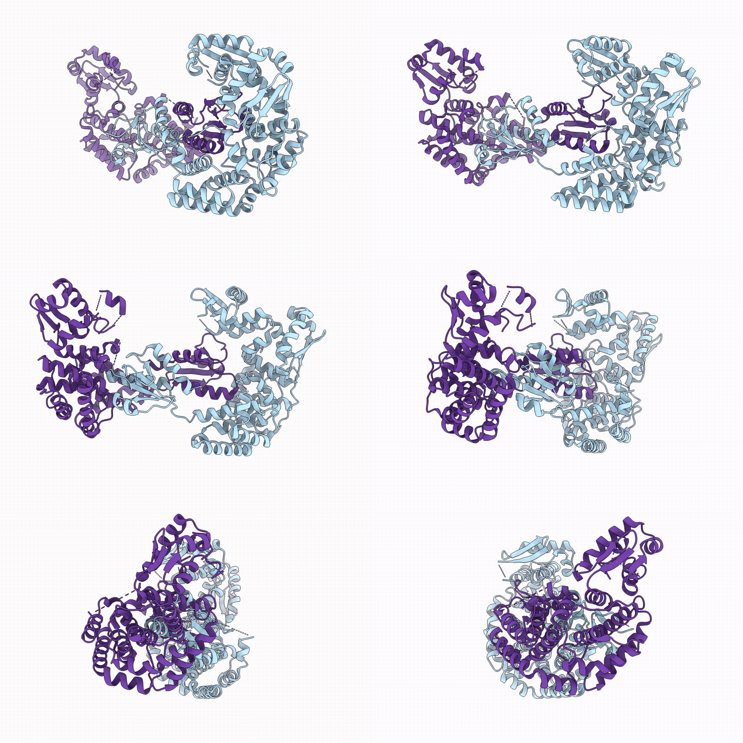
23VJ image
Deposition Date 2026-02-20
Release Date 2026-04-29
Last Version Date 2026-04-29
Entry Detail
PDB ID:
23VJ
Keywords:
Title:
Crystal structure of the MafR protein from Enterococcus faecalis
Biological Source:
Source Organism(s):
Expression System(s):
Method Details:
Experimental Method:
Resolution:
2.30 Å
R-Value Free:
0.24
R-Value Work:
0.21
R-Value Observed:
0.21
Space Group:
P 1 21 1

Macromolecular Entities
Structures with similar UniProt ID
Protein Blast
Polymer Type:polypeptide(L)
Molecule:M protein trans-acting positi
Gene (Uniprot):EGW70_07280
Chain IDs:A, B
Chain Length:488
Number of Molecules:2
Biological Source:Enterococcus faecalis
Primary Citation
Unique structural features and DNA recognition mechanisms of the PRD-containing virulence regulators MafR and MgaSpn.
Biochem.Biophys.Res.Commun. 814 153625 153625 (2026)
PMID: 41865467 DOI: 10.1016/j.bbrc.2026.153625

Abstact

Phosphotransferase system regulatory domain-containing virulence regulators (PCVRs), including Enterococcus faecalis MafR and Streptococcus pneumoniae Mga-like protein (MgaSpn), are transcription factors in pathogenic bacteria that control the transcription of virulence genes by binding dsDNA in promoter regions. Although PCVRs play critical roles in bacterial pathogenesis, the structural and molecular basis by which PCVRs recognize dsDNA for transcriptional regulation has remained unclear. Here, we present the unique structure of MafR and demonstrate the critical role of its two N-terminal helix-turn-helix (HTH) domains in dsDNA binding. MafR adopts a five-domain architecture with an open-ring conformation and dimerizes into a table-like structure, in which the C-terminal domain mediates dimerization in the middle of the dimer structure, and the two N-terminal HTH domains project from one face of the dimer. Our extensive modeling, biochemical, and mutational analyses of the MgaSpn-dsDNA interaction reveal that the tandem HTH domains are required for dsDNA binding by inserting their recognition helices into the major groove of dsDNA. Our findings highlight a unique mode of DNA recognition by PCVRs, in which two HTH domains are simultaneously employed for DNA binding, rather than the single HTH domain typically used by conventional HTH-containing transcriptional regulators.

Legend

Protein

Chemical

Disease

Primary Citation of related structures
Feedback Form
Name
Email
Institute
Feedback