

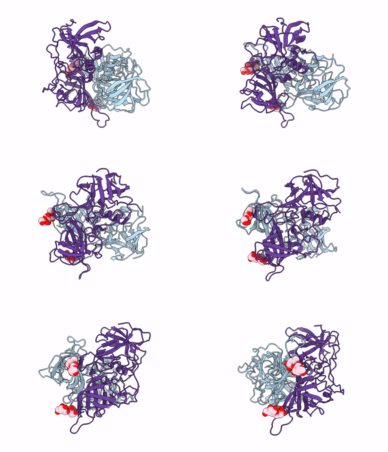
22XH image
Deposition Date 2026-01-27
Release Date 2026-02-25
Last Version Date 2026-04-01
Entry Detail
PDB ID:
22XH
Keywords:
Title:
GII.23/24/25 noroviruses recognize glycans via a conventional glycan-binding site
Biological Source:
Source Organism(s):
Norovirus GII (Taxon ID: 122929)
Expression System(s):
Method Details:
Experimental Method:
Resolution:
1.60 Å
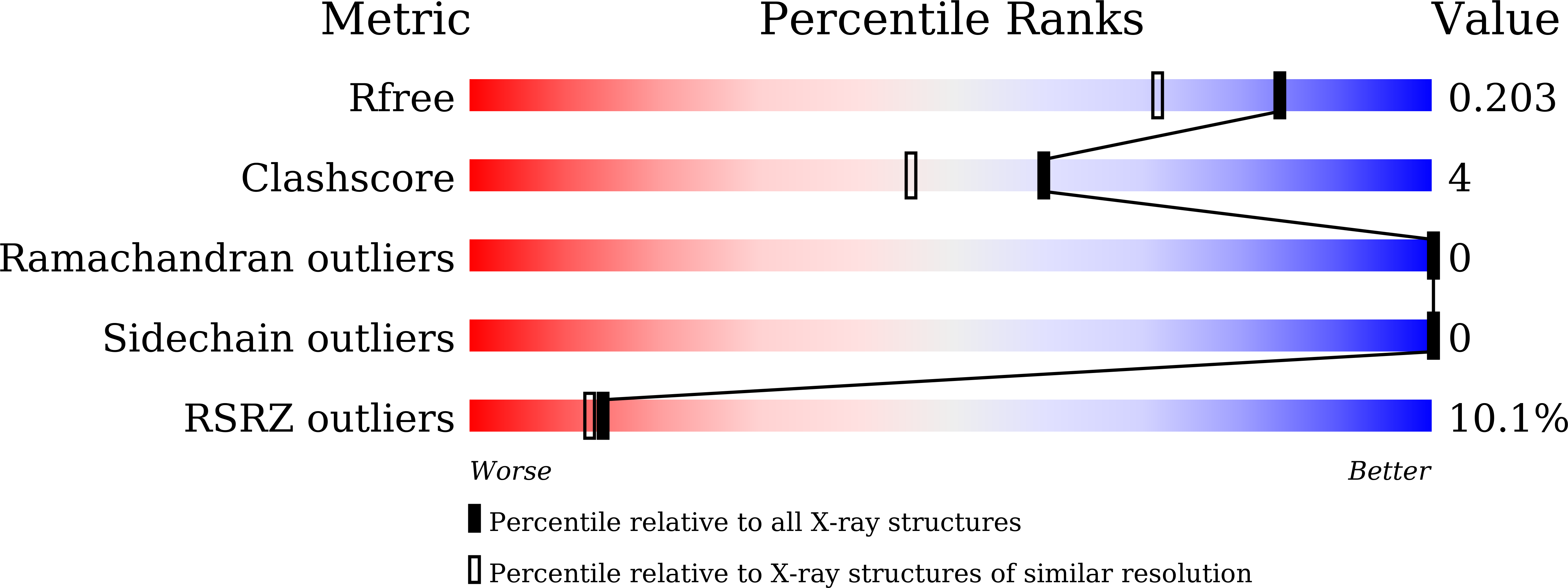
R-Value Free:
0.19
R-Value Work:
0.18
Space Group:
C 1 2 1
Macromolecular Entities
Structures with similar UniProt ID
Protein Blast
Polymer Type:polypeptide(L)
Molecule:VP1
Chain IDs:A, B
Chain Length:307
Number of Molecules:2
Biological Source:Norovirus GII
Primary Citation
GII.23/24/25 noroviruses recognize glycans via a conventional glycan-binding site.
Front Microbiol 17 1767002 1767002 (2026)
PMID: 41853721 DOI: 10.3389/fmicb.2026.1767002

Abstact

INTRODUCTION: Human noroviruses (HuNoVs) are genetically diverse RNA viruses that cause acute gastroenteritis, with genogroup II (GII) accounting for over 90% of global infections. Glycans, particularly histo-blood group antigens (HBGAs), have been identified as attachment factors or receptors for HuNoVs infection. However, the glycan-binding receptors of the later-identified GII genotypes GII.23/24/25 remain elusive. METHODS: We used saliva- and glycan-based ELISA assays to identify the binding spectra of GII.23/24/25 strains. We also solved the crystal structures of their P domains, including the GII.25 P domain in complex with the H disaccharide. Single-point mutagenesis was performed to identify key residues involved in glycan binding. RESULTS: The P domains of GII.24 and GII.25 can recognize multiple types of saliva samples, including both A/B/O secretor and nonsecretor individuals. In contrast, GII.23 primarily binds to B secretor saliva samples. Furthermore, GII.23/24 P domains are able to interact with the H disaccharide, whereas GII.25 exhibits binding affinity for both H disaccharide and B trisaccharide. Crystal structures of GII.23/24/25 P domains revealed high structural similarity, and the complex of GII.25 P domains with H disaccharide was resolved. Single-point mutagenesis identified N352, R353, D382, G443, G444, and H445 as critical residues for H disaccharide binding in GII.25 P domain, while A351 determines glycan-binding specificity. DISCUSSION: Our findings demonstrate that GII.23/24/25 exhibit glycan-binding patterns similar to most other GII HuNoV genotypes. The structural insights provide a better understanding of virus-host evolution and inform the development of therapeutic strategies against HuNoVs.

Legend

Protein

Chemical

Disease

Primary Citation of related structures
Feedback Form
Name
Email
Institute
Feedback