

22HI image
Deposition Date 2026-01-11
Release Date 2026-05-27
Last Version Date 2026-05-27
Entry Detail
PDB ID:
22HI
Title:
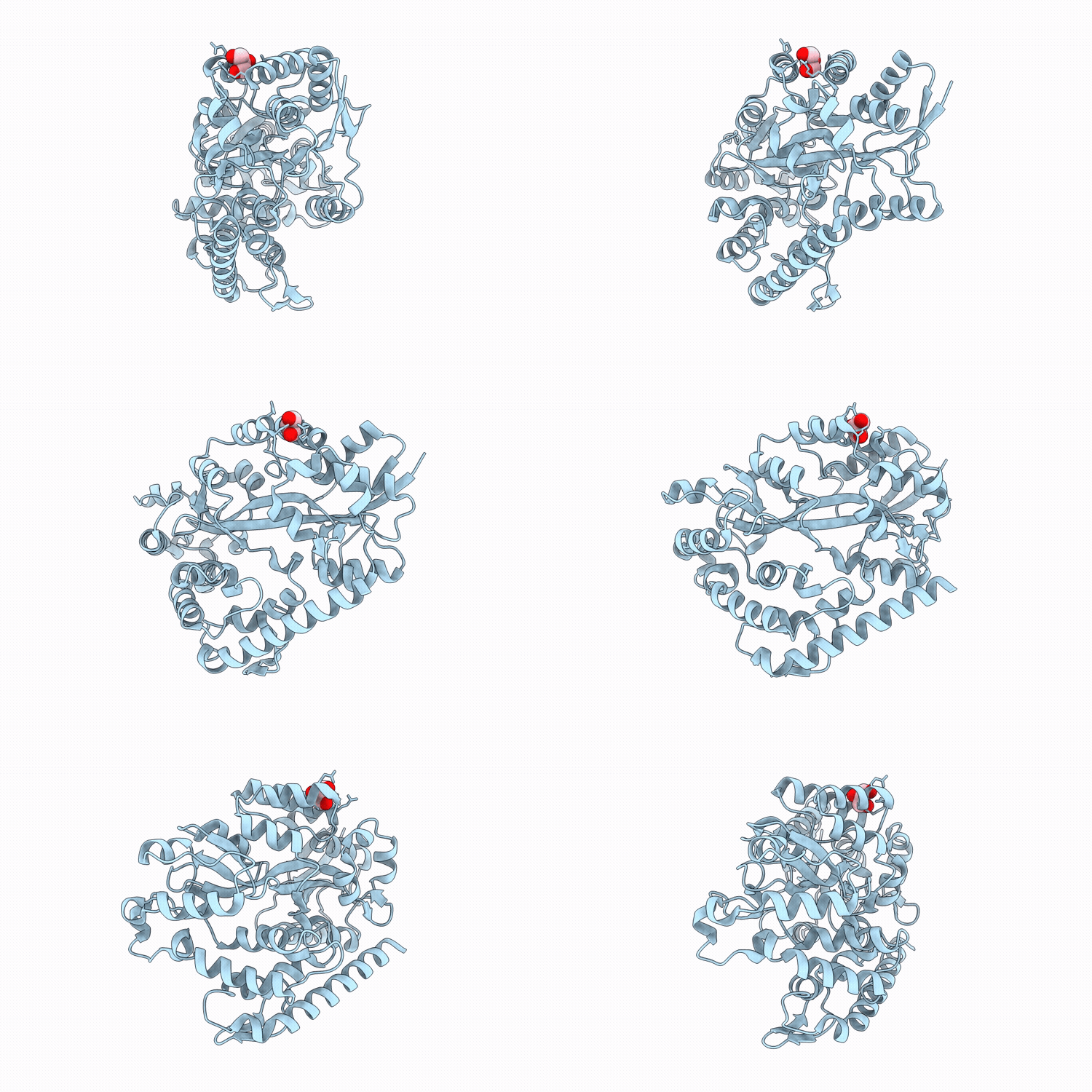
Beta-1,2-glucan-binding protein complexed with cyclic beta-1,2-glucoheptadecaose
Biological Source:
Source Organism(s):
Expression System(s):
Method Details:
Experimental Method:
Resolution:
1.95 Å
R-Value Free:
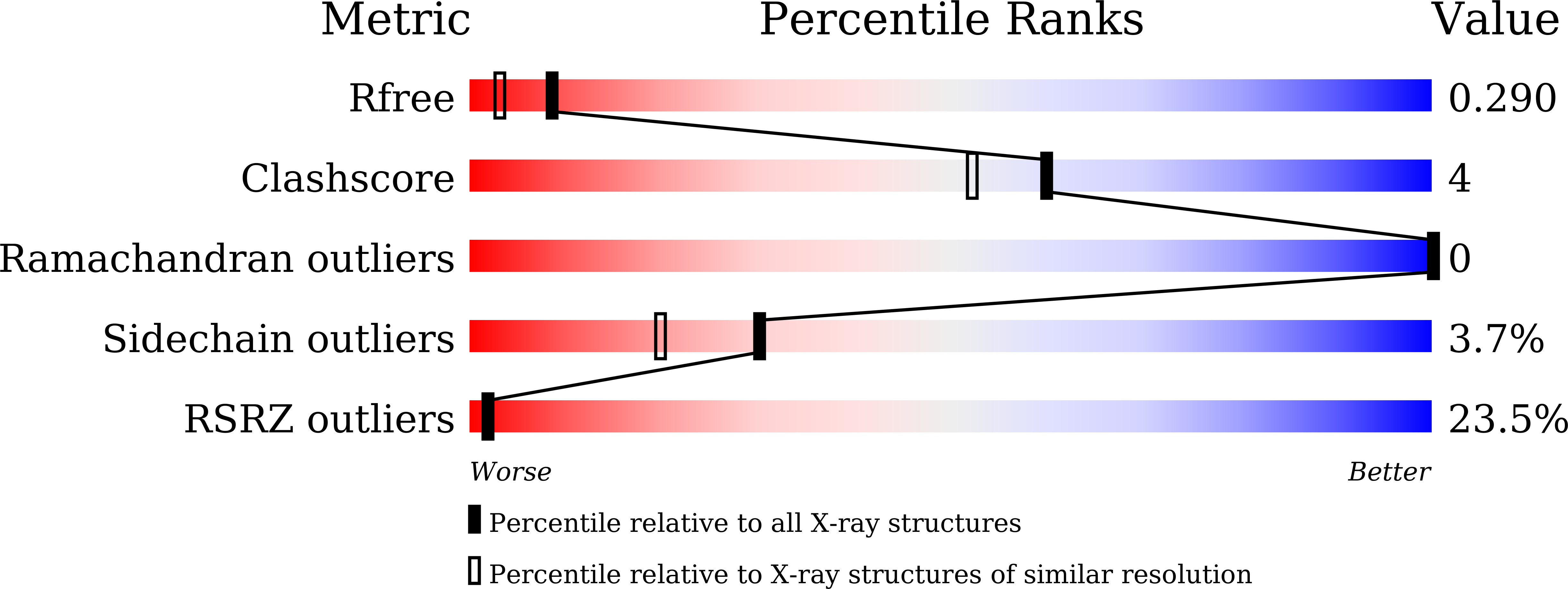
0.28
R-Value Work:
0.24
Space Group:
P 21 21 21
Macromolecular Entities
Structures with similar UniProt ID
Protein Blast
Polymer Type:polypeptide(L)
Molecule:Extracellular solute-binding
Gene (Uniprot):Caur_3857
Chain IDs:A
Chain Length:474
Number of Molecules:1
Biological Source:Chloroflexus aurantiacus Y-400-fl
Ligand Molecules
Primary Citation
Structural and thermodynamic analyses of a novel beta-1,2-glucan binding mode in the ABC transporter solute-binding protein Chy400_4166 from Chloroflexus aurantiacus.
Febs J. ? ? ? (2026)
PMID: 42108234 DOI: 10.1111/febs.70576

Abstact

beta-1,2-Glucans are glucose polymers widely distributed in nature and play various physiological roles in the interactions between organisms such as pathogenicity and symbiosis. While various beta-1,2-glucan-degrading enzymes have been identified recently, transporters incorporating beta-1,2-glucans are still poorly characterized. In this study, we have found a beta-1,2-glucan binding protein of ABC transporter from Chloroflexus aurantiacus Y-400-fl, a filamentous anoxygenic phototrophic bacterium. The protein showed a clear affinity for linear beta-1,2-glucan in the gel shift assay. Isothermal titration calorimetric analysis revealed high binding affinities for both linear and cyclic beta-1,2-glucans, unlike for the barley beta-glucan. The recorded binding constants were high for the binding of the ABC transporter to beta-1,2-glucans. The observed unfavorable negative entropy change may have resulted from conformational restraints upon complex formation. Complex structures with linear beta-1,2-glucan and cyclic beta-1,2-glucans with degrees of polymerization of 17-20 were obtained using X-ray crystallography. Ten glucose units, designated A-J from the nonreducing end, were shared among the substrates in the complexes. Unit G is recognized by W74, W308, and D336, which are highly conserved residues within the phylogenetic group Chy400_4166. The substrate-binding mode of Chy400_4166 is completely different from that of the beta-1,2-glucooligosaccharide-binding protein from Listeria innocua. The discovery of a new type of beta-1,2-glucan-related binding protein has expanded our understanding of the metabolism of beta-1,2-glucans.

Legend

Protein

Chemical

Disease

Primary Citation of related structures
Feedback Form
Name
Email
Institute
Feedback