21ZE image
Deposition Date 2026-01-04
Release Date 2026-04-22
Last Version Date 2026-04-29
Entry Detail
PDB ID:
21ZE
Title:
Crystal structure of the petrobactin-binding protein FatB from Bacillus cereus complexed with ferric petrobactin
Biological Source:
Source Organism(s):
Expression System(s):
Method Details:
Experimental Method:
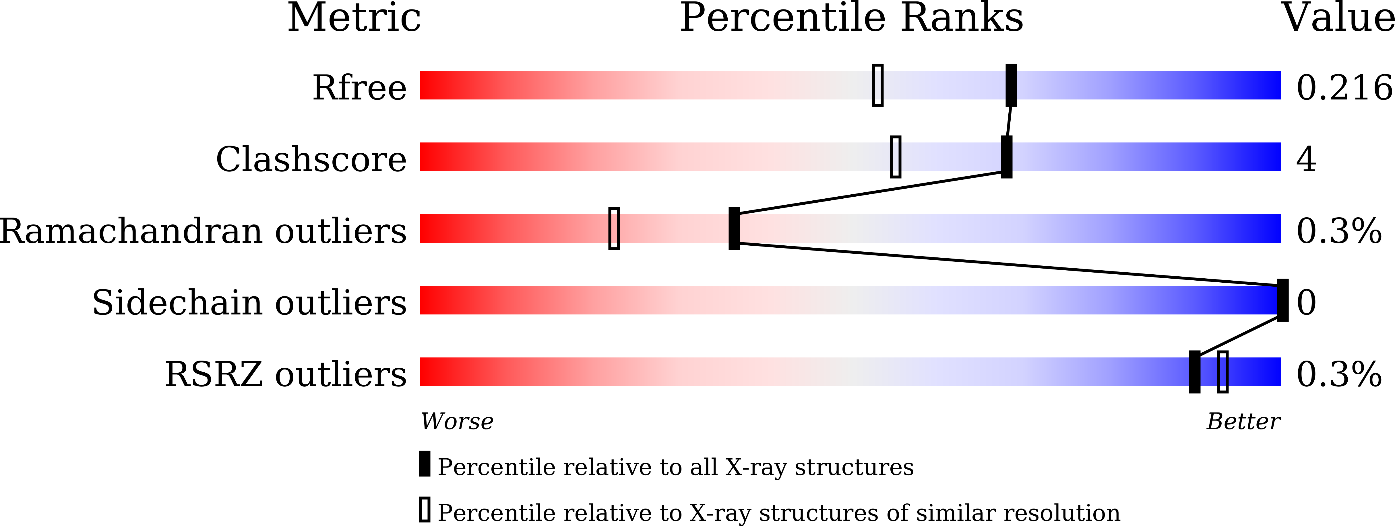
Resolution:
1.78 Å
R-Value Free:
0.21
R-Value Work:
0.16
R-Value Observed:
0.17
Space Group:
P 1 21 1
Macromolecular Entities
Structures with similar UniProt ID
Protein Blast
Polymer Type:polypeptide(L)
Molecule:Ferric anguibactin-binding pr
Gene (Uniprot):BC_5106
Chain IDs:A
Chain Length:302
Number of Molecules:1
Biological Source:Bacillus cereus ATCC 14579
Primary Citation
Structural basis of FatB-mediated iron uptake via tyrosine/histidine direct coordination accompanying long-distance domain reorganization.
Nat Commun ? ? ? (2026)
PMID: 42000734 DOI: 10.1038/s41467-026-72127-y

Abstact

Iron is an essential cofactor for fundamental biological processes. However, Fe(III) is poorly soluble under aerobic conditions, limiting its bioavailability. To secure this essential nutrient, bacteria release high-affinity siderophores that capture environmental Fe(III) and are subsequently imported into the cell as ferric siderophore complexes. While biochemical studies have characterized siderophore uptake in Bacillus species, atomic-level mechanisms of recognition and coordination remain unclear. Here, we investigate the siderophore-binding protein FatB from Bacillus cereus and its interactions with its siderophore, petrobactin (PB), as well as with ferric petrobactin (FePB) and its ferric photoproduct (FePB(nu)). Crystal structures of apo- and ferric-ligand-bound FatB, supported by biophysical and mutational analyses, reveal that ferric-siderophore binding induces substantial domain closure of FatB. This conformational transition involves an extensive ~29-A reorganization of a flexible loop, which positions His252 alongside Tyr317 to directly coordinate the Fe(III) center in the FePB-FatB complex. This protein-derived coordination mode is maintained in the FePB(nu)-FatB complex, where a structured water network preserves interfacial complementarity and functional recognition. These findings provide a structural framework for siderophore recognition and iron acquisition and illustrate how active-site coordination and domain reorganization facilitate robust capture of chemically labile ligands, offering insights for antimicrobial development targeting bacterial iron uptake.

Legend

Protein

Chemical

Disease

Primary Citation of related structures
Feedback Form
Name
Email
Institute
Feedback