

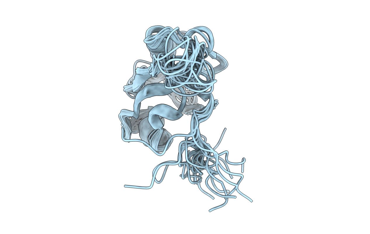
1mc7 image
Deposition Date 2002-08-05
Release Date 2003-09-23
Last Version Date 2024-05-22
Entry Detail
PDB ID:
1MC7
Title:
Solution Structure of mDvl1 PDZ domain
Biological Source:
Source Organism(s):
Mus musculus (Taxon ID: 10090)
Expression System(s):
Method Details:
Experimental Method:
Conformers Calculated:
320
Conformers Submitted:
20
Selection Criteria:
target function

Macromolecular Entities
Structures with similar UniProt ID
Protein Blast
Polymer Type:polypeptide(L)
Molecule:Segment polarity protein dish
Gene (Uniprot):Dvl1
Mutagens:C88A
Chain IDs:A
Chain Length:95
Number of Molecules:1
Biological Source:Mus musculus
Ligand Molecules
Primary Citation
Direct binding of the PDZ domain of Dishevelled to a conserved internal sequence in the C-terminal region of Frizzled
Mol. Cell 12 1251 1260 (2003)
PMID: 14636582 DOI: 10.1016/S1097-2765(03)00427-1

Abstact

The cytoplasmic protein Dishevelled (Dvl) and the associated membrane-bound receptor Frizzled (Fz) are essential in canonical and noncanonical Wnt signaling pathways. However, the molecular mechanisms underlying this signaling are not well understood. By using NMR spectroscopy, we determined that an internal sequence of Fz binds to the conventional peptide binding site in the PDZ domain of Dvl; this type of site typically binds to C-terminal binding motifs. The C-terminal region of the Dvl inhibitor Dapper (Dpr) and Frodo bound to the same site. In Xenopus, Dvl binding peptides of Fz and Dpr/Frodo inhibited canonical Wnt signaling and blocked Wnt-induced secondary axis formation in a dose-dependent manner, but did not block noncanonical Wnt signaling mediated by the DEP domain. Together, our results identify a missing molecular connection within the Wnt pathway. Differences in the binding affinity of the Dvl PDZ domain and its binding partners may be important in regulating signal transduction by Dvl.

Legend

Protein

Chemical

Disease

Primary Citation of related structures
Feedback Form
Name
Email
Institute
Feedback