

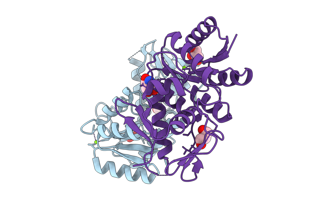
1YS7 image
Deposition Date 2005-02-07
Release Date 2006-02-07
Last Version Date 2023-10-25
Entry Detail
PDB ID:
1YS7
Title:
Crystal structure of the response regulator protein prrA complexed with Mg2+
Biological Source:
Source Organism(s):
Expression System(s):
Method Details:
Experimental Method:
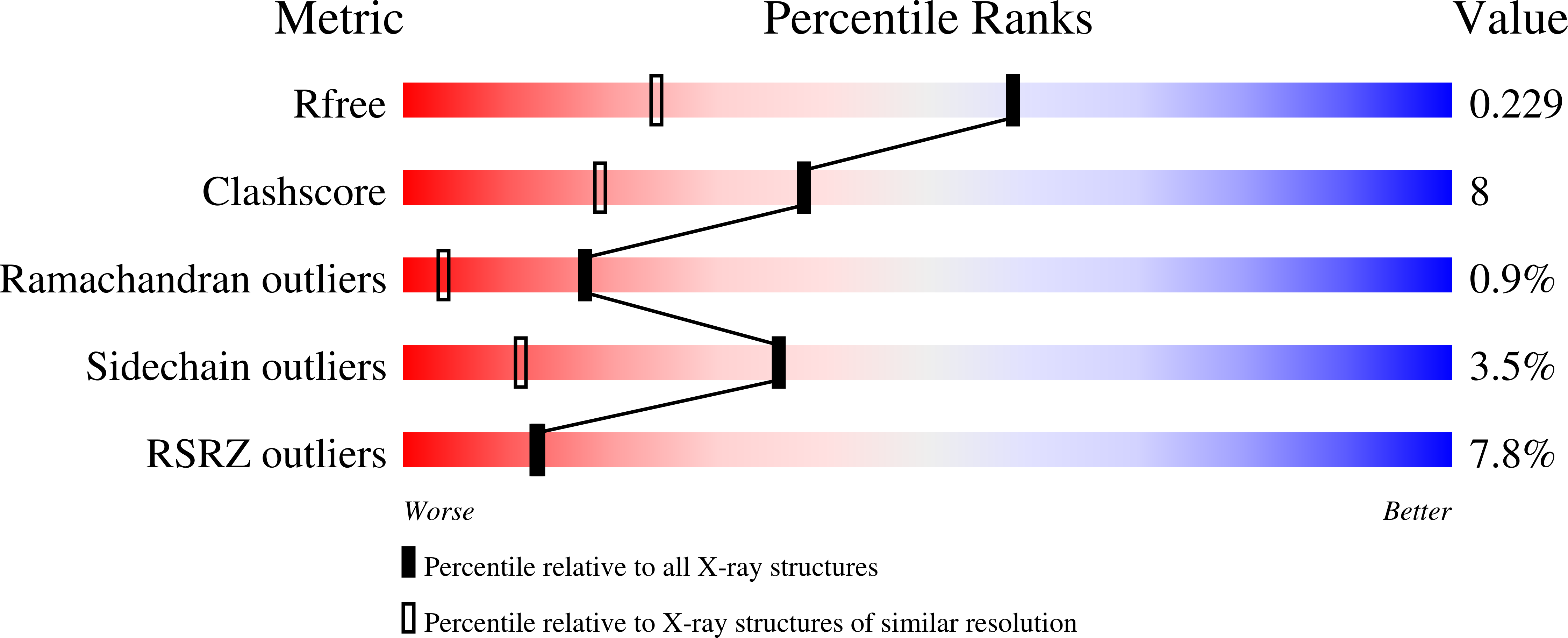
Resolution:
1.58 Å
R-Value Free:
0.22
R-Value Work:
0.18
R-Value Observed:
0.18
Space Group:
P 1
Macromolecular Entities
Structures with similar UniProt ID
Protein Blast
Polymer Type:polypeptide(L)
Molecule:Transcriptional regulatory pr
Chain IDs:A, B
Chain Length:233
Number of Molecules:2
Biological Source:Mycobacterium tuberculosis
Primary Citation
The structural basis of signal transduction for the response regulator PrrA from Mycobacterium tuberculosis.
J. Biol. Chem. 281 9659 9666 (2006)
PMID: 16434396 DOI: 10.1074/jbc.M512004200

Abstact

The structure of the two-domain response regulator PrrA from Mycobacterium tuberculosis shows a compact structure in the crystal with a well defined interdomain interface. The interface, which does not include the interdomain linker, makes the recognition helix and the trans-activation loop of the effector domain inaccessible for interaction with DNA. Part of the interface involves hydrogen-bonding interactions of a tyrosine residue in the receiver domain that is believed to be involved in signal transduction, which, if disrupted, would destabilize the interdomain interface, allowing a more extended conformation of the molecule, which would in turn allow access to the recognition helix. In solution, there is evidence for an equilibrium between compact and extended forms of the protein that is far toward the compact form when the protein is inactivated but moves toward a more extended form when activated by the cognate sensor kinase PrrB.

Legend

Protein

Chemical

Disease

Primary Citation of related structures
Feedback Form
Name
Email
Institute
Feedback