

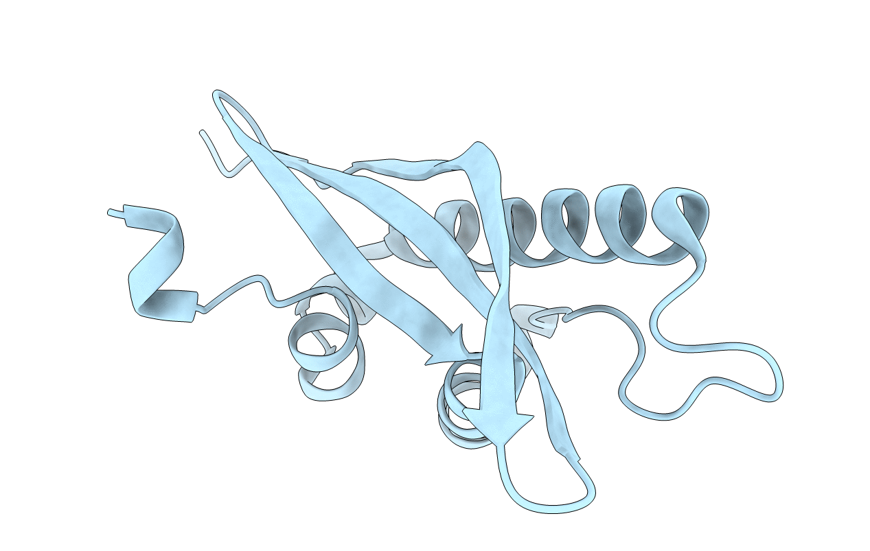
1XTE image
Deposition Date 2004-10-21
Release Date 2004-11-16
Last Version Date 2024-02-14
Entry Detail
PDB ID:
1XTE
Keywords:
Title:
crystal structure of CISK-PX domain
Biological Source:
Source Organism(s):
Mus musculus (Taxon ID: 10090)
Expression System(s):
Method Details:
Experimental Method:
Resolution:
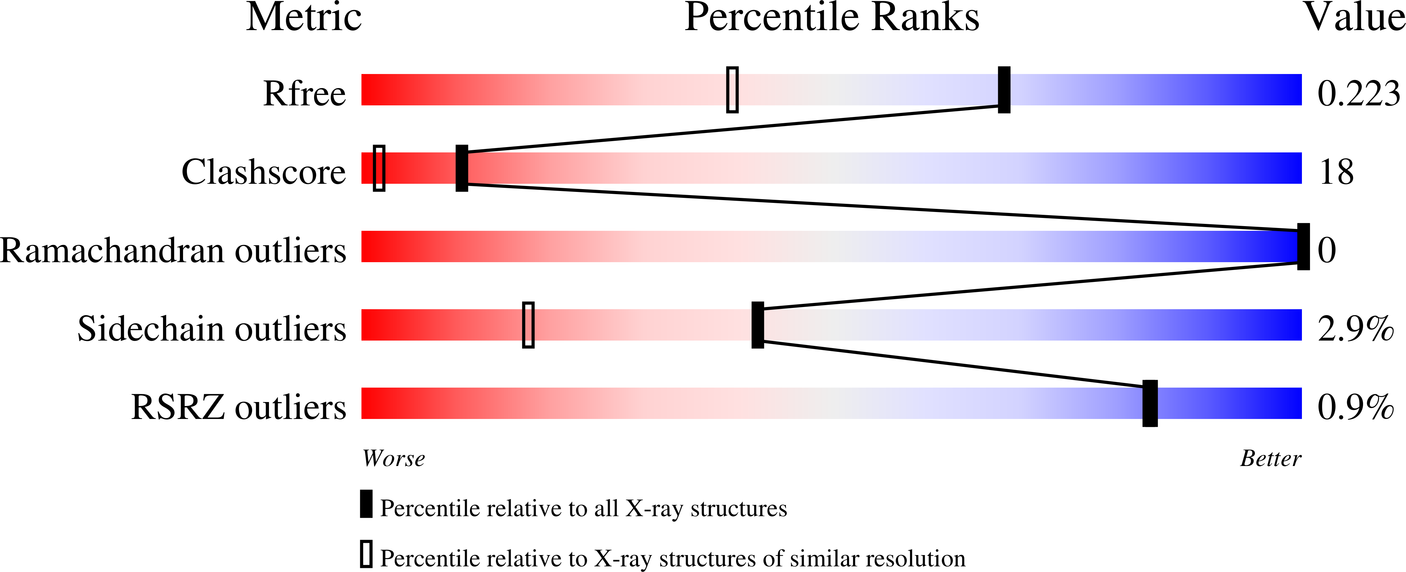
1.60 Å
R-Value Free:
0.22
R-Value Work:
0.18
R-Value Observed:
0.18
Space Group:
I 4
Macromolecular Entities
Structures with similar UniProt ID
Protein Blast
Polymer Type:polypeptide(L)
Molecule:Serine/threonine-protein kina
Gene (Uniprot):Sgk3
Chain IDs:A
Chain Length:154
Number of Molecules:1
Biological Source:Mus musculus
Primary Citation
Structural basis of membrane targeting by the Phox homology domain of cytokine-independent survival kinase (CISK-PX)
J. Biol. Chem. 279 30662 30669 (2004)
PMID: 15126499 DOI: 10.1074/jbc.M404107200

Abstact

The cytokine-independent survival kinase (CISK) in the serum and glucocorticoid-regulated kinase family plays an important role in mediating cell growth and survival. N-terminal to its catalytic kinase domain, CISK contains a phox homology (PX) domain, a phosphoinositide-binding motif that directs the membrane localization of CISK and regulates CISK activity. We have determined the crystal structures of the mouse CISK-PX domain to unravel the structural basis of membrane targeting of CISK. In addition to the specific interactions conferred by the phosphoinositide-binding pocket, the structure suggests that a hydrophobic loop region and a hydrophilic beta-turn contribute to the interactions with the membrane. Furthermore, biochemical studies reveal that CISK-PX dimerizes in the presence of the linker between the PX domain and kinase domain, suggesting a multivalent mechanism in membrane localization of CISK.

Legend

Protein

Chemical

Disease

Primary Citation of related structures
Feedback Form
Name
Email
Institute
Feedback