

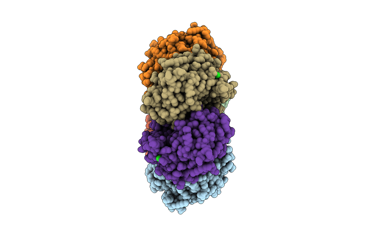
1XE3 image
Deposition Date 2004-09-09
Release Date 2004-10-26
Last Version Date 2023-08-23
Entry Detail
PDB ID:
1XE3
Keywords:
Title:
Crystal Structure of purine nucleoside phosphorylase DeoD from Bacillus anthracis
Biological Source:
Source Organism(s):
Expression System(s):
Method Details:
Experimental Method:
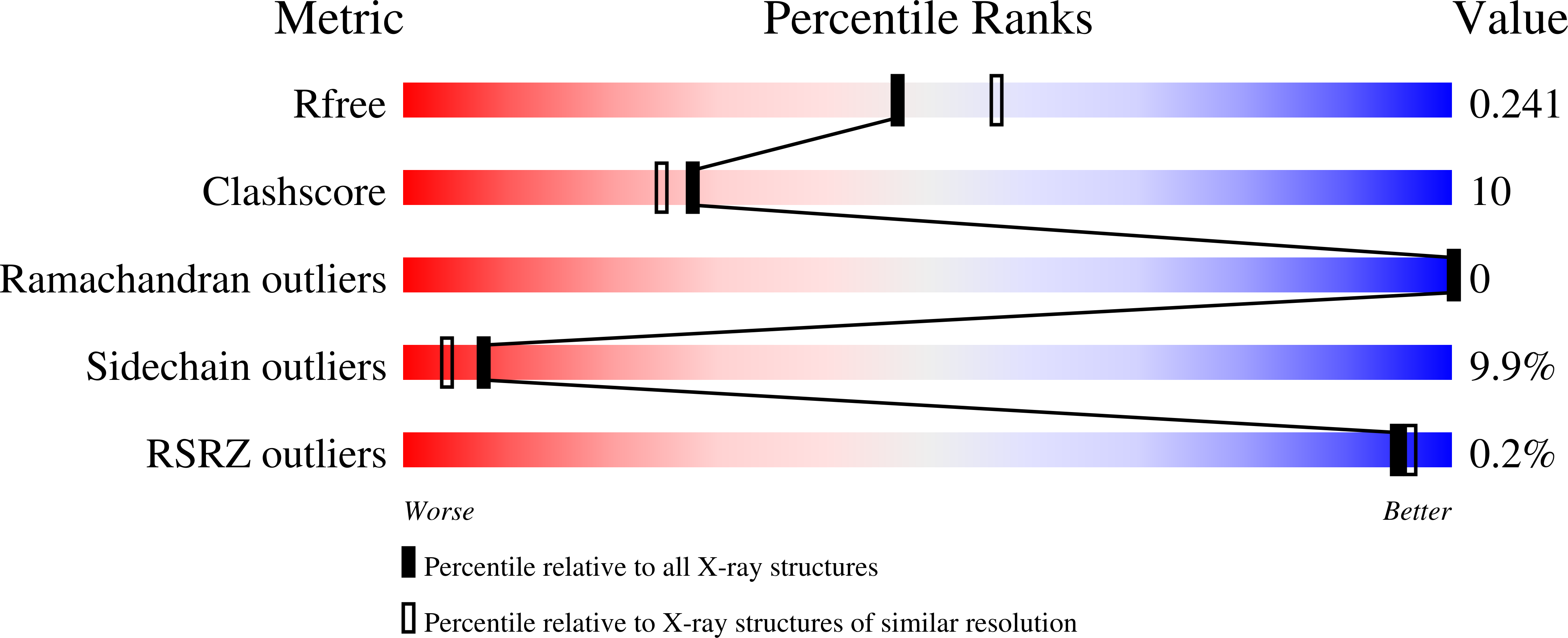
Resolution:
2.24 Å
R-Value Free:
0.23
R-Value Work:
0.18
R-Value Observed:
0.18
Space Group:
P 21 21 21
Macromolecular Entities
Structures with similar UniProt ID
Protein Blast
Polymer Type:polypeptide(L)
Molecule:purine nucleoside phosphoryla
Gene (Uniprot):deoD
Chain IDs:A, B, C, D, E, F
Chain Length:245
Number of Molecules:6
Biological Source:Bacillus anthracis
Ligand Molecules
Primary Citation
Structure of purine nucleoside phosphorylase (DeoD) from Bacillus anthracis.
Acta Crystallogr. Sect. F Struct. Biol. Cryst. Commun. 61 459 462 (2005)
PMID: 16511068 DOI: 10.1107/S174430910501095X

Abstact

Protein structures from the causative agent of anthrax (Bacillus anthracis) are being determined as part of a structural genomics programme. Amongst initial candidates for crystallographic analysis are enzymes involved in nucleotide biosynthesis, since these are recognized as potential targets in antibacterial therapy. Purine nucleoside phosphorylase is a key enzyme in the purine-salvage pathway. The crystal structure of purine nucleoside phosphorylase (DeoD) from B. anthracis has been solved by molecular replacement at 2.24 A resolution and refined to an R factor of 18.4%. This is the first report of a DeoD structure from a Gram-positive bacterium.

Legend

Protein

Chemical

Disease

Primary Citation of related structures
Feedback Form
Name
Email
Institute
Feedback