

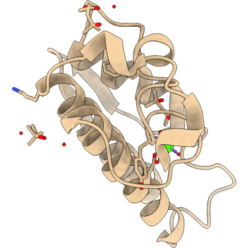
1VKQ image
Deposition Date 2004-06-12
Release Date 2004-08-31
Last Version Date 2024-11-20
Entry Detail
PDB ID:
1VKQ
Keywords:
Title:
A re-determination of the structure of the triple mutant (K53,56,120M) of phospholipase A2 at 1.6A resolution using sulphur-SAS at 1.54A wavelength
Biological Source:
Source Organism(s):
Bos taurus (Taxon ID: 9913)
Expression System(s):
Method Details:
Experimental Method:
Resolution:
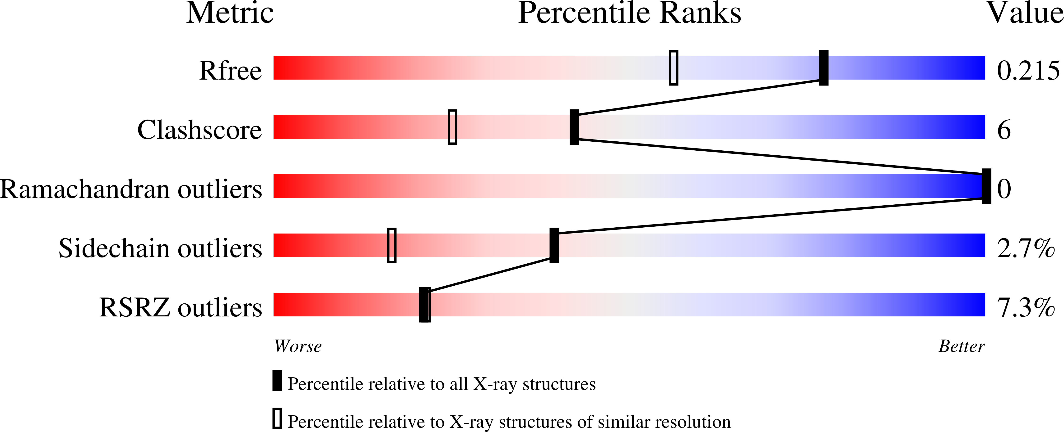
1.60 Å
R-Value Free:
0.21
R-Value Work:
0.17
R-Value Observed:
0.17
Space Group:
P 31 2 1
Macromolecular Entities
Polymer Type:polypeptide(L)
Molecule:Phospholipase A2
Gene (Uniprot):PLA2G1B
Mutagens:K53M, K56M, K120M
Chain IDs:A
Chain Length:123
Number of Molecules:1
Biological Source:Bos taurus
Primary Citation
A redetermination of the structure of the triple mutant (K53,56,120M) of phospholipase A2 at 1.6 A resolution using sulfur-SAS at 1.54 A wavelength.
Acta Crystallogr. D Biol. Crystallogr. 60 1586 1590 (2004)
PMID: 15333929 DOI: 10.1107/S090744490401697X

Abstact

The crystal structure of the triple mutant K53,56,120M of bovine pancreatic phospholipase A(2) has been redetermined using sulfur single-wavelength anomalous scattering. The synchrotron data were collected at lambda = 1.54 A and the crystal diffracted to 1.6 A resolution. The program SOLVE was used to locate the heavy atoms and to estimate the initial phases and the resulting map was then subjected to RESOLVE. The output of 455 non-H atoms, including 12 S atoms, one calcium ion and one chloride ion, were then subjected to ARP/wARP followed by REFMAC. With the improved phases, the automatic model building successfully built more than 85% of the 123 residues, excluding the N- and C-terminal residues. The final crystallographic R factor is 17.7% (R(free) = 21.7%). The refined model consists of 954 non-H protein atoms, 165 water O atoms, three 2-methyl-2,4-pentanediol (MPD) molecules, one calcium ion and one chloride ion. The present work is yet another example that shows the utility of single-wavelength anomalous scattering data for solving a protein structure.

Legend

Protein

Chemical

Disease

Primary Citation of related structures
Feedback Form
Name
Email
Institute
Feedback