

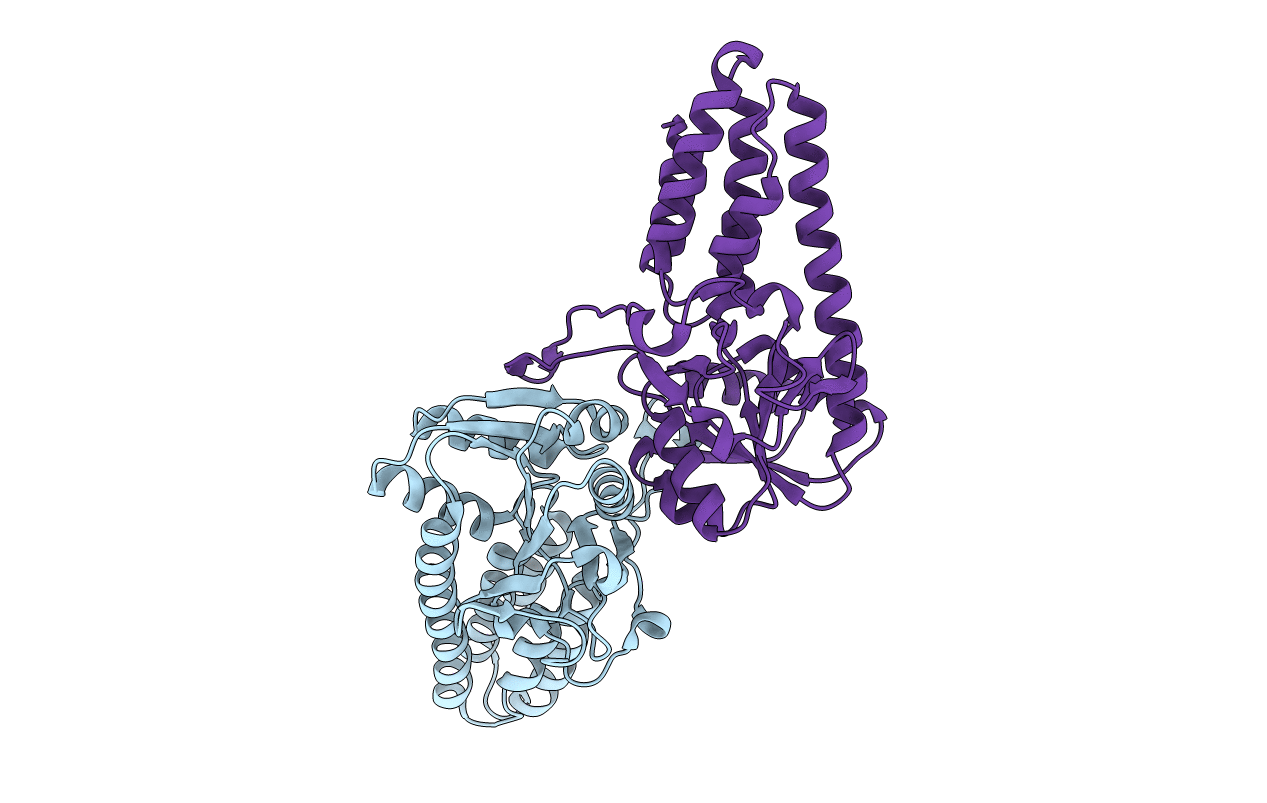
1VB5 image
Deposition Date 2004-02-22
Release Date 2004-12-07
Last Version Date 2024-11-13
Entry Detail
PDB ID:
1VB5
Keywords:
Title:
Crystal Structure Analysis of the Pyrococcus horikoshii OT3 translation initiation factor eIF-2B
Biological Source:
Source Organism(s):
Expression System(s):
Method Details:
Experimental Method:
Resolution:
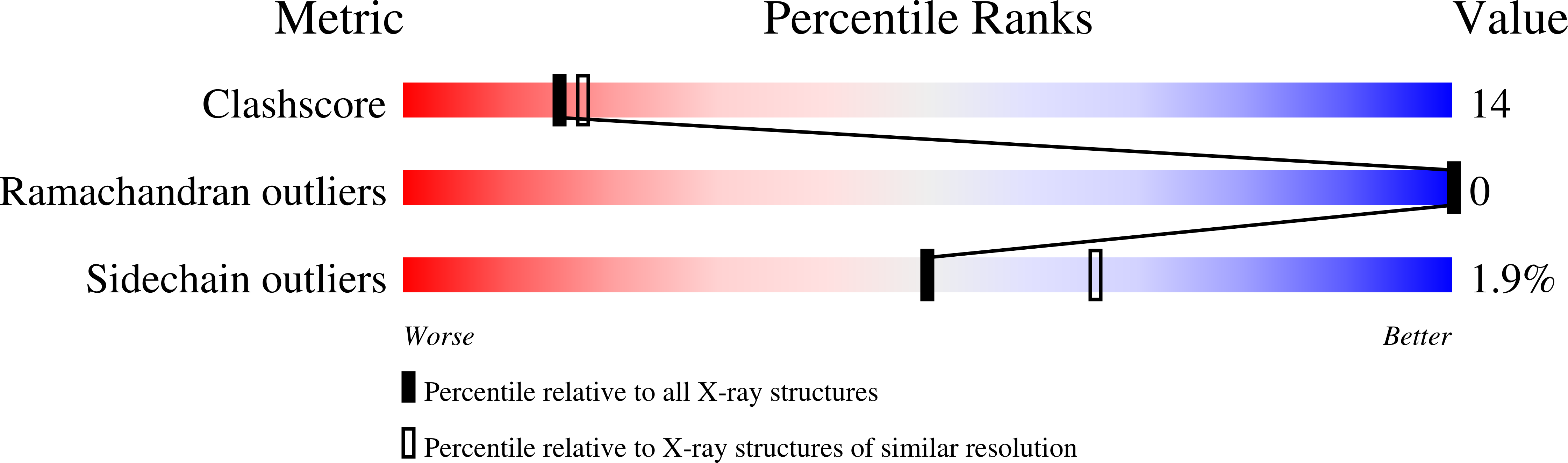
2.20 Å
R-Value Free:
0.23
R-Value Work:
0.20
Space Group:
P 63 2 2
Macromolecular Entities
Polymer Type:polypeptide(L)
Molecule:translation initiation factor
Chain IDs:A, B
Chain Length:276
Number of Molecules:2
Biological Source:Pyrococcus horikoshii
Primary Citation
Crystal structure of the regulatory subunit of archaeal initiation factor 2B (aIF2B) from hyperthermophilic archaeon Pyrococcus horikoshii OT3: a proposed structure of the regulatory subcomplex of eukaryotic IF2B
Biochem.Biophys.Res.Commun. 319 725 732 (2004)
PMID: 15184043 DOI: 10.1016/j.bbrc.2004.05.045

Abstact

Eukaryotic translation initiation factor 2B (eIF2B) is the guanine-nucleotide exchange factor for eukaryotic initiation factor 2 (eIF2). eIF2B is a heteropentameric protein composed of alpha- subunits. The alpha, beta, and delta subunits form a regulatory subcomplex, while the gamma and form a catalytic subcomplex. Archaea possess homologues of alpha, beta, and delta subunits of eIF2B. Here, we report the three-dimensional structure of an archaeal regulatory subunit (aIF2Balpha) from the hyperthermophilic archaeon Pyrococcus horikoshii OT3 determined by X-ray crystallography at 2.2A resolution. aIF2Balpha consists of two subdomains, an N-domain (residues 1-95) and a C-domain (residues 96-276), connected by a long alpha-helix (alpha5: 78-106). The N-domain contains a five helix bundle structure, while the C-domain folds into the alpha/beta structure, thus showing similarity to D-ribose-5-phosphate isomerase structure. The presence of two molecules in the crystallographic asymmetric unit and the gel filtration analysis suggest a dimeric structure of aIF2Balpha in solution, interacting with each other by C-domains. Furthermore, the crystallographic 3-fold symmetry generates a homohexameric structure of aIF2Balpha; the interaction is primarily mediated by the long alpha-helix at the N-domains. This structure suggests an architecture of the three subunits, alpha, beta, and delta, in the regulatory subcomplex within eIF2B.

Legend

Protein

Chemical

Disease

Primary Citation of related structures
Feedback Form
Name
Email
Institute
Feedback