Deposition Date
2003-12-04
Release Date
2004-09-24
Last Version Date
2024-05-15
Entry Detail
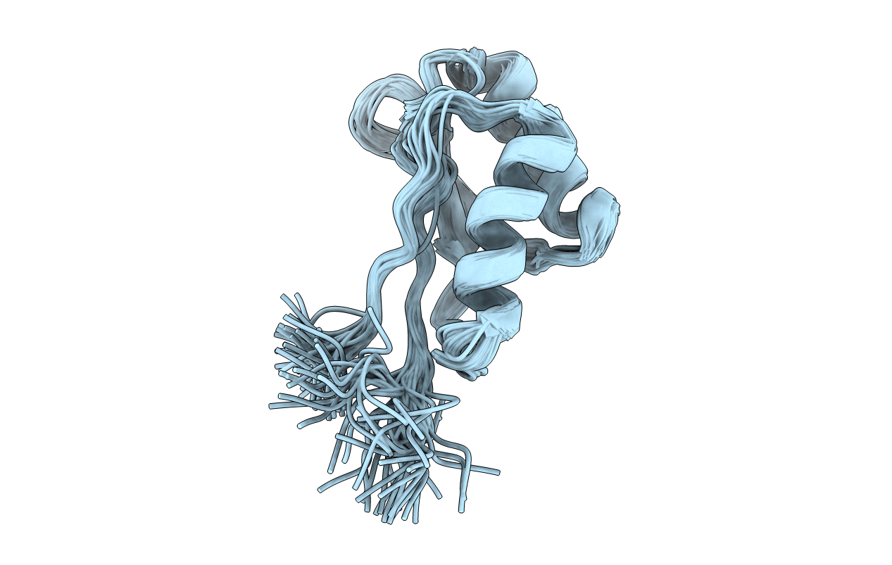
PDB ID:
1UTA
Keywords:
Title:
Solution structure of the C-terminal RNP domain from the divisome protein FtsN
Biological Source:
Source Organism(s):
ESCHERICHIA COLI (Taxon ID: 562)
Expression System(s):
Method Details:
Experimental Method:
Conformers Calculated:
50
Conformers Submitted:
45
Selection Criteria:
LOW NOE ENERGY