

1TXY image
Deposition Date 2004-07-06
Release Date 2004-11-16
Last Version Date 2024-11-06
Entry Detail
PDB ID:
1TXY
Title:
E. coli PriB
Biological Source:
Source Organism(s):
Escherichia coli (Taxon ID: 562)
Expression System(s):
Method Details:
Experimental Method:
Resolution:
2.00 Å
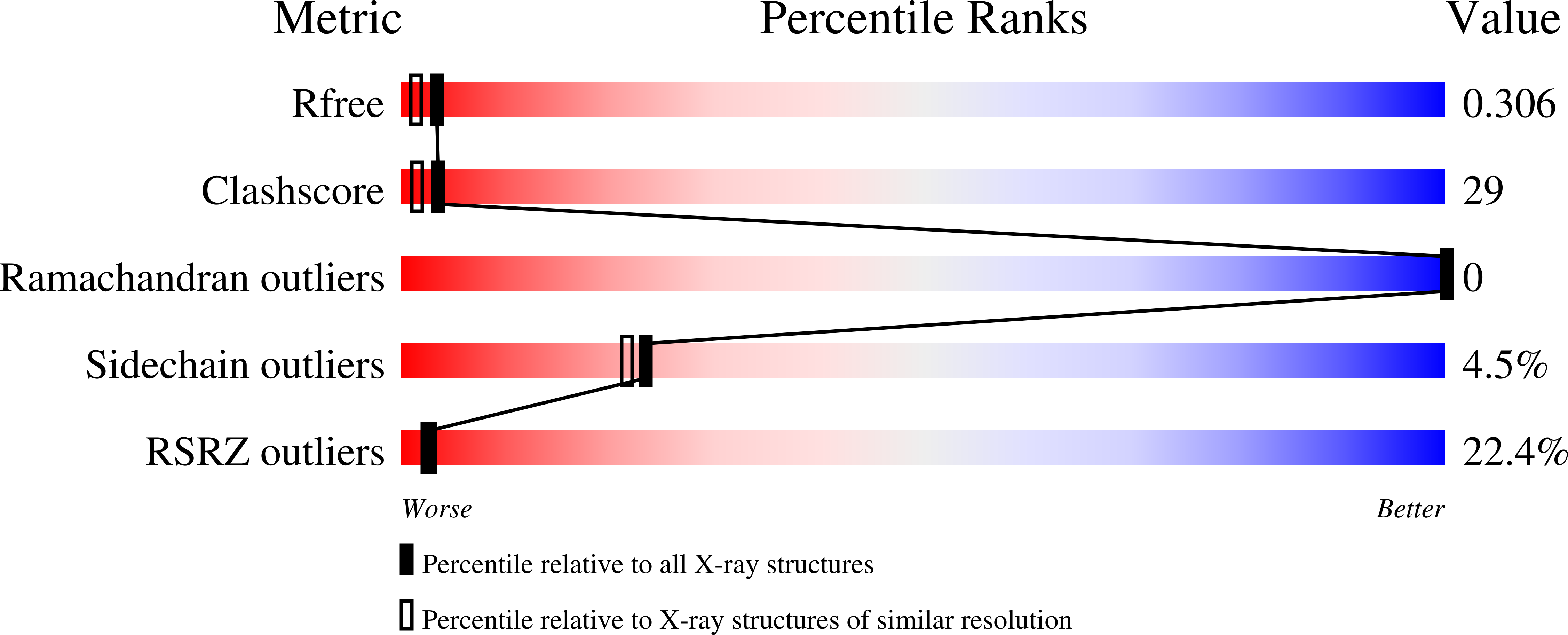
R-Value Free:
0.28
R-Value Work:
0.25
R-Value Observed:
0.26
Space Group:
P 21 21 21
Macromolecular Entities
Structures with similar UniProt ID
Protein Blast
Polymer Type:polypeptide(L)
Molecule:Primosomal replication protei
Gene (Uniprot):priB
Chain IDs:A, B
Chain Length:104
Number of Molecules:2
Biological Source:Escherichia coli
Modified Residue
Compound ID Chain ID Parent Comp ID Details 2D Image
MSE A MET SELENOMETHIONINE
Primary Citation
Crystal structure of PriB, a component of the Escherichia coli replication restart primosome
Structure 12 1967 1975 (2004)
PMID: 15530361 DOI: 10.1016/j.str.2004.09.004

Abstact

Maintenance of genome stability following DNA damage requires origin-independent reinitiation of DNA replication at repaired replication forks. In E. coli, PriA, PriB, PriC, and DnaT play critical roles in recognizing repaired replication forks and reloading the replisome onto the template to reinitiate DNA replication. Here, we report the 2.0 A resolution crystal structure of E. coli PriB, revealing a dimer that consists of a single structural domain formed by two oligonucleotide/oligosaccharide binding (OB) folds. Structural similarity of PriB to single-stranded DNA binding proteins reveals insights into its mechanisms of DNA binding. The structure further establishes a putative protein interaction surface that may contribute to the role of PriB in primosome assembly by facilitating interactions with PriA and DnaT. This is the first high-resolution structure of a protein involved in oriC-independent replisome loading and provides unique insight into mechanisms of replication restart in E. coli.

Legend

Protein

Chemical

Disease

Primary Citation of related structures
Feedback Form
Name
Email
Institute
Feedback