

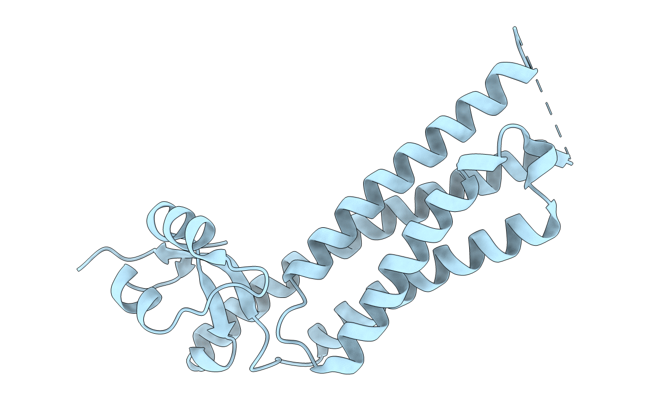
1SZI image
Deposition Date 2004-04-05
Release Date 2004-07-27
Last Version Date 2024-02-14
Entry Detail
PDB ID:
1SZI
Title:
Crystal Structure of the C-terminus of TIP47
Biological Source:
Source Organism(s):
Mus musculus (Taxon ID: 10090)
Expression System(s):
Method Details:
Experimental Method:
Resolution:
2.80 Å
R-Value Free:
0.27
R-Value Work:
0.23
R-Value Observed:
0.23
Space Group:
P 63 2 2

Macromolecular Entities
Polymer Type:polypeptide(L)
Molecule:mannose-6-phosphate receptor
Gene (Uniprot):Plin3
Chain IDs:A
Chain Length:247
Number of Molecules:1
Biological Source:Mus musculus
Ligand Molecules
Primary Citation
Structure of a Lipid Droplet Protein: The PAT Family Member TIP47
Structure 12 1199 1207 (2004)
PMID: 15242596 DOI: 10.1016/j.str.2004.04.021

Abstact

The perilipin/ADRP/TIP47 (PAT) proteins localize to the surface of intracellular neutral lipid droplets. Perilipin is essential for lipid storage and hormone regulated lipolysis in adipocytes, and perilipin null mice exhibit a dramatic reduction in adipocyte lipid stores. A significant fraction of the approximately 200 amino acid N-terminal region of the PAT proteins consists of 11-mer helical repeats that are also found in apolipoproteins and other lipid-associated proteins. The C-terminal 60% of TIP47, a representative PAT protein, comprises a monomeric and independently folded unit. The crystal structure of the C-terminal portion of TIP47 was determined and refined at 2.8 A resolution. The structure consists of an alpha/beta domain of novel topology and a four-helix bundle resembling the LDL receptor binding domain of apolipoprotein E. The structure suggests an analogy between PAT proteins and apolipoproteins in which helical repeats interact with lipid while the ordered C-terminal region is involved in protein:protein interactions.

Legend

Protein

Chemical

Disease

Primary Citation of related structures
Feedback Form
Name
Email
Institute
Feedback