

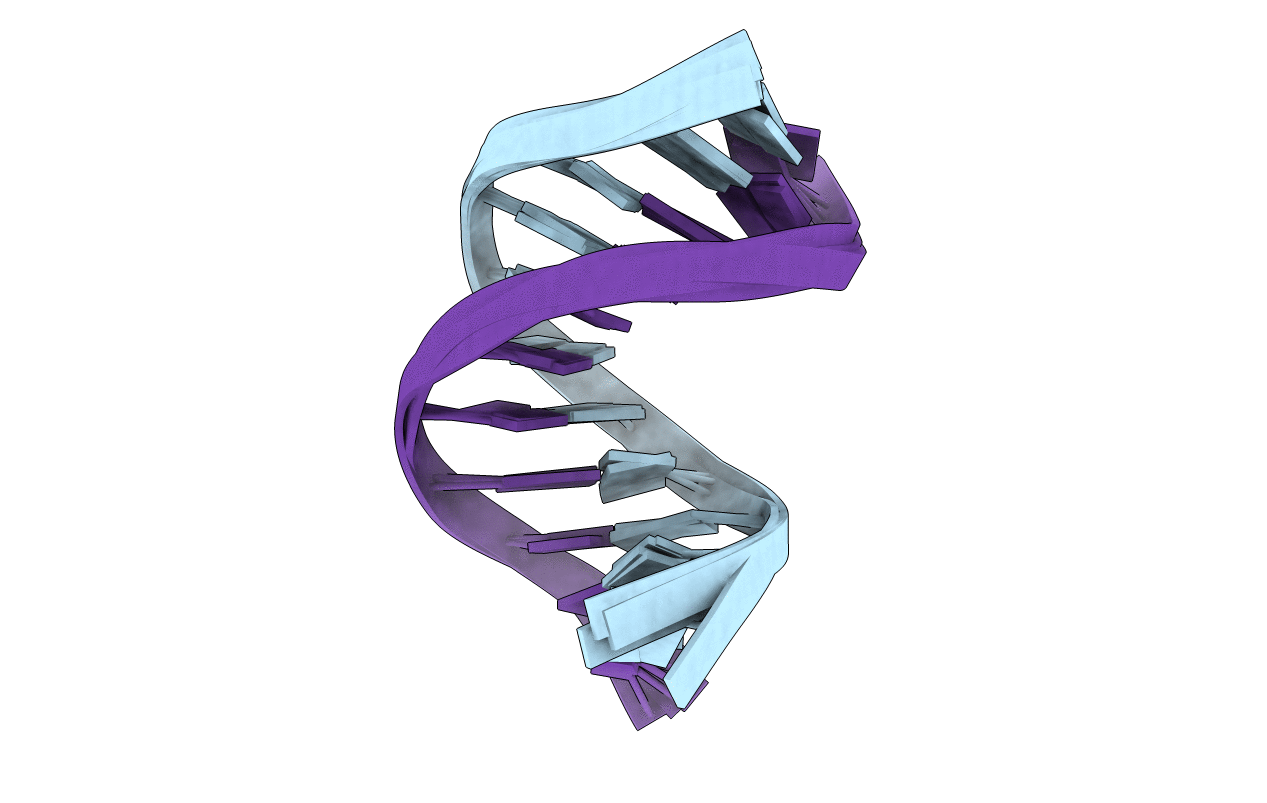
1SKP image
Deposition Date 1998-05-20
Release Date 1999-01-13
Last Version Date 2024-05-22
Entry Detail
PDB ID:
1SKP
Keywords:
Title:
NMR STRUCTURE OF D(GCATATGATAG)(DOT)D(CTATCATATGC): A CONSENSUS SEQUENCE FOR PROMOTERS RECOGNIZED BY SIGMA-K RNA POLYMERASE, 4 STRUCTURES
Method Details:
Experimental Method:
Conformers Calculated:
20
Conformers Submitted:
4
Selection Criteria:
CONVERGENCE, R FACTORS, ENERGIES

Macromolecular Entities
Polymer Type:polydeoxyribonucleotide
Molecule:SIGMA-K RNA POLYMERASE CONSEN
Chain IDs:A
Chain Length:11
Number of Molecules:1
Biological Source:
Polymer Type:polydeoxyribonucleotide
Molecule:SIGMA-K RNA POLYMERASE CONSEN
Chain IDs:B
Chain Length:11
Number of Molecules:1
Biological Source:
Ligand Molecules
Primary Citation
Nuclear magnetic resonance structure of d(GCATATGATAG). d(CTATCATATGC): a consensus sequence for promoters recognized by sigma K RNA polymerase.
Biochemistry 37 11745 11761 (1998)
PMID: 9718297 DOI: 10.1021/bi980481n

Abstact

The three-dimensional structure of d(GCATATGATAG).d(CTATCATATGC), from the promoter region of a gene regulating sporulation in Bacillus subtilis mother cells, was determined utilizing two-dimensional nuclear Overhauser effect (2D NOE) and double-quantum-filtered COSY (2QF-COSY) spectra. To minimize the effect of methods used to obtain restraints and refine structure, several variables were studied. Interproton distance bounds were calculated very conservatively by running the complete relaxation matrix program MARDIGRAS hundreds of times using 2D NOE spectra for exchangeable and for nonexchangeable protons at different mixing times, assuming different overall correlation times and different starting structures. The 435 distance restraints were used with two different structural refinement methods: restrained molecular dynamics (rMD) and restrained Monte Carlo calculations (rMC). Refinement using different procedures and starting structures resulted in essentially the same structure (<0.8 A rmsd), indicating that the structure is defined by experimental restraints and not the refinement method or variables used. R factors indicate the structures fit the experimental NOE data very well. Some helical parameters, notably large negative X displacement, are characteristic of A-DNA, but others are characteristic of B-DNA. As with TG.CA steps in other duplex DNA sequences studied in our laboratory, the two TG.CA steps have a positive roll, with T6-G7 exhibiting the largest, and consequently a bent helix axis. The converged structure represents a time-averaged structure. However, multiple conformations, especially in deoxyriboses, were evident from vicinal coupling constants obtained from quantitative simulations of 2QF-COSY cross-peaks and from persistent inconsistencies in experimental distances due to nonlinear conformational averaging.

Legend

Protein

Chemical

Disease

Primary Citation of related structures
Feedback Form
Name
Email
Institute
Feedback