

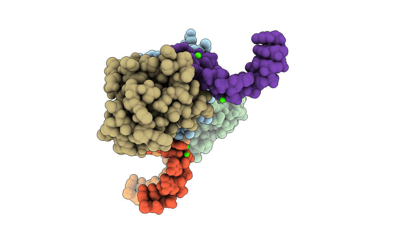
1SDS image
Deposition Date 2004-02-13
Release Date 2004-05-18
Last Version Date 2024-02-14
Entry Detail
PDB ID:
1SDS
Title:
Structure of protein L7Ae bound to a K-turn derived from an archaeal box H/ACA sRNA
Biological Source:
Source Organism(s):
Expression System(s):
Method Details:
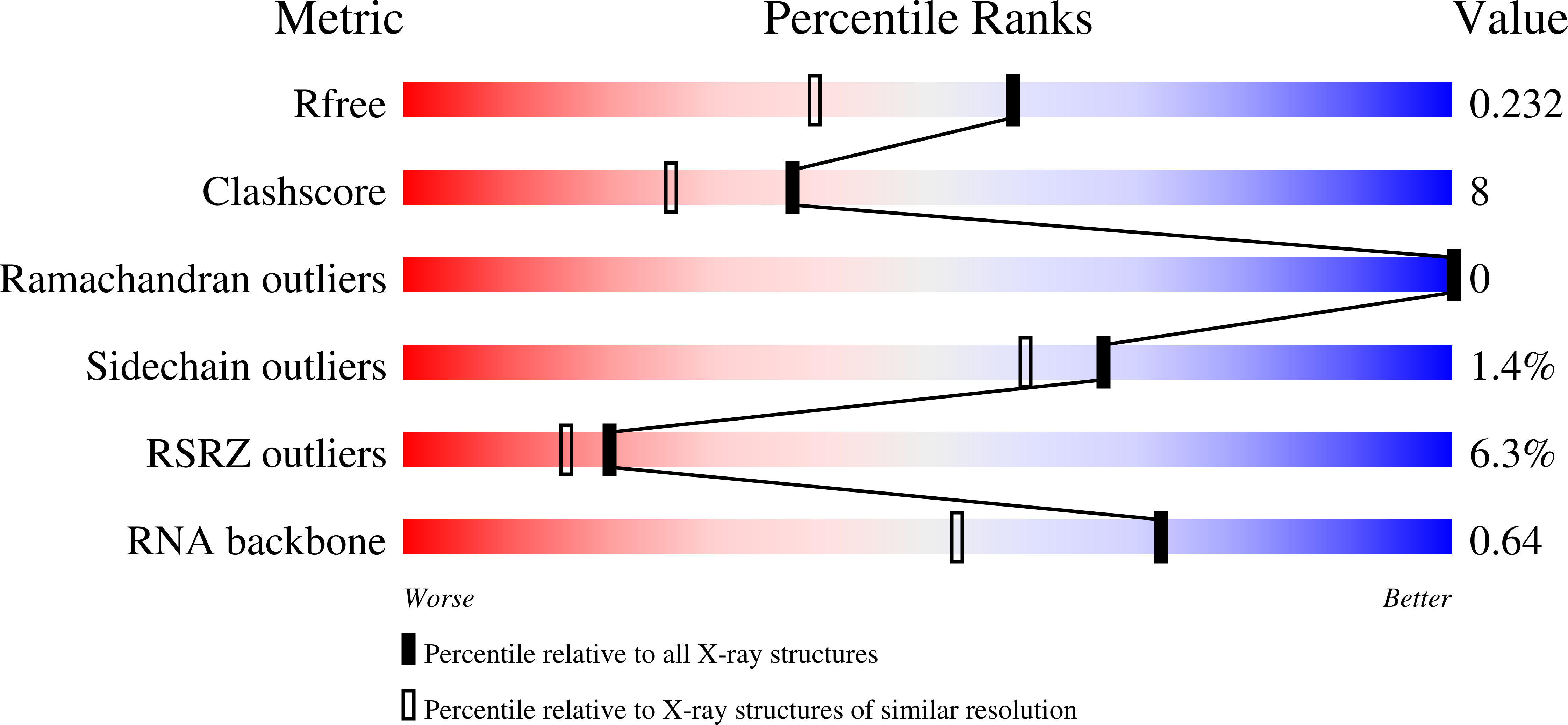
Experimental Method:
Resolution:
1.80 Å
R-Value Free:
0.22
R-Value Work:
0.20
R-Value Observed:
0.20
Space Group:
F 2 2 2
Macromolecular Entities
Structures with similar UniProt ID
Protein Blast
Polymer Type:polypeptide(L)
Molecule:50S ribosomal protein L7Ae
Gene (Uniprot):rpl7ae
Chain IDs:D (auth: A), E (auth: B), F (auth: C)
Chain Length:117
Number of Molecules:3
Biological Source:Methanocaldococcus jannaschii
Primary Citation
Structure of Protein L7Ae Bound to a K-Turn Derived from an Archaeal Box H/ACA sRNA at 1.8 A Resolution.
Structure 12 893 903 (2004)
PMID: 15130481 DOI: 10.1016/j.str.2004.03.015

Abstact

The archaeal RNA binding protein L7Ae and its eukaryotic homolog 15.5 kDa/Snu13 recognize K-turns. This structural motif is canonically comprised of two stems (one with tandem A.G base pairs, the other with Watson-Crick pairs) linked by an asymmetric internal loop. L7Ae recognizes conventional K-turns in ribosomal and box C/D RNAs but also binds specifically to some box H/ACA RNAs at terminal stem loops. These have the A.G paired stem, but lack the Watson-Crick stem. The structure of Methanococcus jannaschii L7Ae bound to a symmetric duplex RNA without Watson-Crick stems demonstrates how a binding site for this component of diverse ribonucleoprotein complexes can be constructed with only the A.G stem and the loop. The RNA adopts a functional conformation with the aid of a base triple and tight binding of divalent cations. Comparison with the 15.5 kDa/Snu13-RNA complex structure suggests why the eukaryotic homolog does not recognize terminal stem loop L7Ae binding sites.

Legend

Protein

Chemical

Disease

Primary Citation of related structures
Feedback Form
Name
Email
Institute
Feedback