

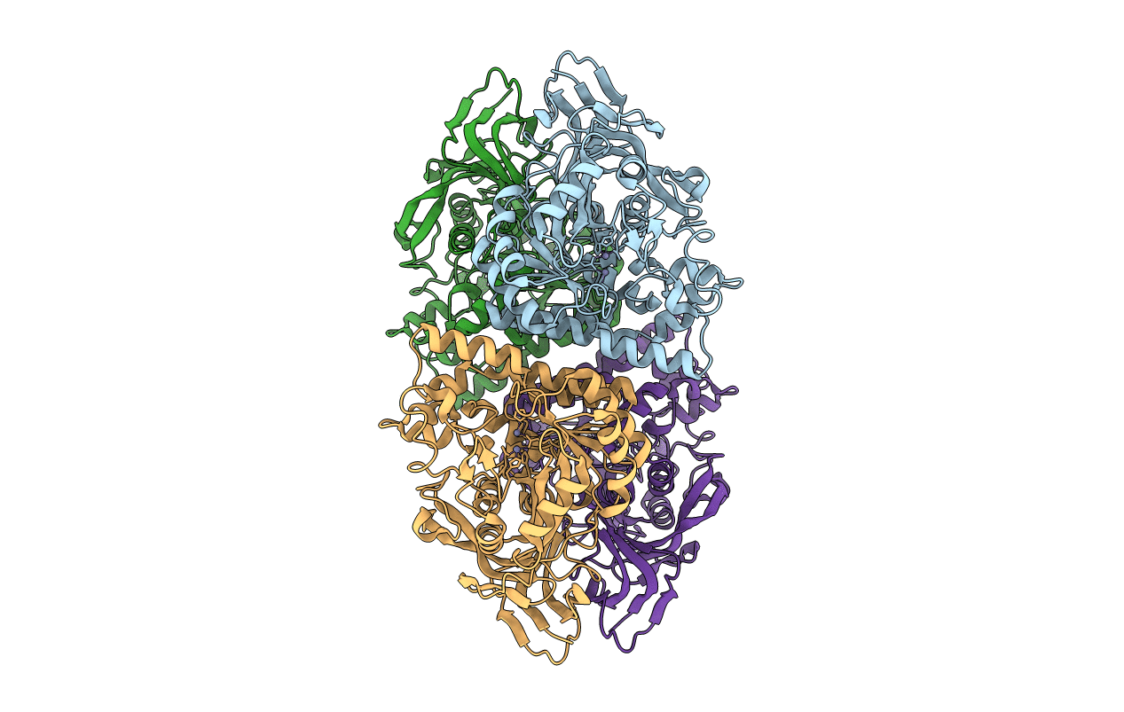
1NFG image
Deposition Date 2002-12-14
Release Date 2003-07-15
Last Version Date 2023-11-15
Entry Detail
PDB ID:
1NFG
Keywords:
Title:
Structure of D-hydantoinase
Biological Source:
Source Organism(s):
Expression System(s):
Method Details:
Experimental Method:
Resolution:
2.70 Å
R-Value Free:
0.23
R-Value Work:
0.22
R-Value Observed:
0.22
Space Group:
P 1 21 1

Macromolecular Entities
Polymer Type:polypeptide(L)
Molecule:D-hydantoinase
Gene (Uniprot):hyuA
Chain IDs:A, B, C, D
Chain Length:457
Number of Molecules:4
Biological Source:Ralstonia pickettii
Modified Residue
Compound ID Chain ID Parent Comp ID Details 2D Image
KCX A LYS LYSINE NZ-CARBOXYLIC ACID
Ligand Molecules
Primary Citation
Crystal Structure of D-Hydantoinase from Burkholderia pickettii at a Resolution of 2.7 Angstroms: Insights into the Molecular Basis of Enzyme Thermostability.
J. Bacteriol. 185 4038 4049 (2003)
PMID: 12837777 DOI: 10.1128/JB.185.14.4038-4049.2003

Abstact

D-Hydantoinase (D-HYD) is an industrial enzyme that is widely used in the production of D-amino acids which are precursors for semisynthesis of antibiotics, peptides, and pesticides. This report describes the crystal structure of D-hydantoinase from Burkholderia pickettii (HYD(Bp)) at a 2.7-A resolution. The structure of HYD(Bp) consists of a core (alpha/beta)(8) triose phosphate isomerase barrel fold and a beta-sheet domain, and the catalytic active site consists of two metal ions and six highly conserved amino acid residues. Although HYD(Bp) shares only moderate sequence similarity with D-HYDs from Thermus sp. (HYD(Tsp)) and Bacillus stearothermophilus (HYD(Bst)), whose structures have recently been solved, the overall structure and the structure of the catalytic active site are strikingly similar. Nevertheless, the amino acids that compose the substrate-binding site are less conserved and have different properties, which might dictate the substrate specificity. Structural comparison has revealed insights into the molecular basis of the differential thermostability of D-HYDs. The more thermostable HYD(Tsp) contains more aromatic residues in the interior of the structure than HYD(Bp) and HYD(Bst). Changes of large aromatic residues in HYD(Tsp) to smaller residues in HYD(Bp) or HYD(Bst) decrease the hydrophobicity and create cavities inside the structure. HYD(Tsp) has more salt bridges and hydrogen-bonding interactions and less oxidation susceptible Met and Cys residues on the protein surface than HYD(Bp) and HYD(Bst). Besides, HYD(Tsp) also contains more rigid Pro residues. These factors are likely to make major contributions to the varying thermostability of these enzymes. This information could be exploited in helping to engineer more thermostable mesophilic enzymes.

Legend

Protein

Chemical

Disease

Primary Citation of related structures
Feedback Form
Name
Email
Institute
Feedback