

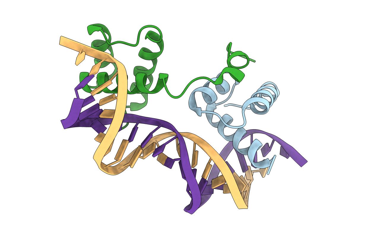
1LE8 image
Deposition Date 2002-04-09
Release Date 2002-05-03
Last Version Date 2024-02-14
Entry Detail
PDB ID:
1LE8
Title:
Crystal Structure of the MATa1/MATalpha2-3A Heterodimer Bound to DNA Complex
Biological Source:
Source Organism(s):
Expression System(s):
Method Details:
Experimental Method:
Resolution:
2.30 Å
R-Value Free:
0.29
R-Value Work:
0.25
Space Group:
P 65

Macromolecular Entities
Polymer Type:polypeptide(L)
Molecule:MATING-TYPE PROTEIN A-1
Chain IDs:C (auth: A)
Chain Length:53
Number of Molecules:1
Biological Source:
Polymer Type:polypeptide(L)
Molecule:Mating-type protein alpha-2
Mutagens:S181A, N182A, R185A
Chain IDs:D (auth: B)
Chain Length:83
Number of Molecules:1
Biological Source:Saccharomyces cerevisiae
Primary Citation
Structural and Thermodynamic Characterization of the DNA Binding Properties of a Triple Alanine Mutant of MATalpha2
Structure 10 961 971 (2002)
PMID: 12121651 DOI: 10.1016/S0969-2126(02)00790-6

Abstact

Triply mutated MATalpha2 protein, alpha2-3A, in which all three major groove-contacting residues are mutated to alanine, is defective in binding DNA alone or in complex with Mcm1 yet binds with MATa1 with near wild-type affinity and specificity. To gain insight into this unexpected behavior, we determined the crystal structure of the a1/alpha2-3A/DNA complex. The structure shows that the triple mutation causes a collapse of the alpha2-3A/DNA interface that results in a reorganized set of alpha2-3A/DNA contacts, thereby enabling the mutant protein to recognize the wild-type DNA sequence. Isothermal titration calorimetry measurements reveal that a much more favorable entropic component stabilizes the a1/alpha2-3A/DNA complex than the alpha2-3A/DNA complex. The combined structural and thermodynamic studies provide an explanation of how partner proteins influence the sequence specificity of a DNA binding protein.

Legend

Protein

Chemical

Disease

Primary Citation of related structures
Feedback Form
Name
Email
Institute
Feedback