

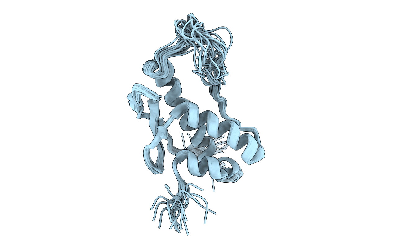
1KMD image
Deposition Date 2001-12-14
Release Date 2002-06-12
Last Version Date 2024-05-22
Entry Detail
PDB ID:
1KMD
Title:
SOLUTION STRUCTURE OF THE VAM7P PX DOMAIN
Biological Source:
Source Organism(s):
Expression System(s):
Method Details:
Experimental Method:
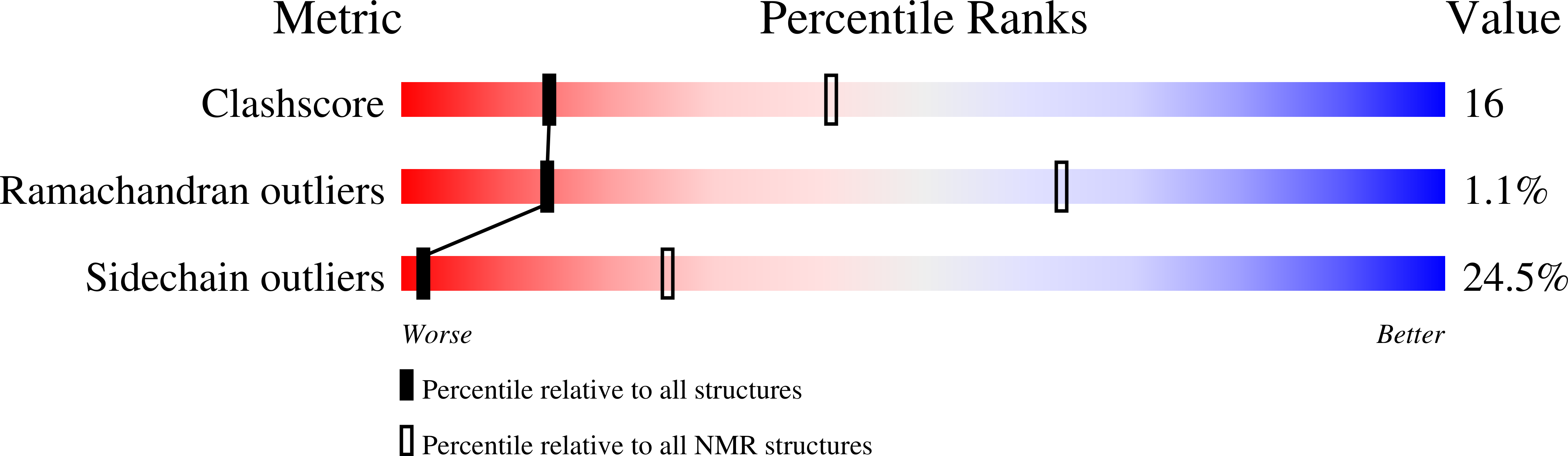
Conformers Calculated:
500
Conformers Submitted:
20
Selection Criteria:
STRUCTURES WITH THE MINIMAL NOE ENERGY
Macromolecular Entities
Polymer Type:polypeptide(L)
Molecule:Vacuolar morphogenesis protei
Gene (Uniprot):VAM7
Chain IDs:A
Chain Length:117
Number of Molecules:1
Biological Source:Saccharomyces cerevisiae
Ligand Molecules
Primary Citation
Solution structure of the Vam7p PX domain.
Biochemistry 41 5956 5962 (2002)
PMID: 11993989 DOI: 10.1021/bi020050b

Abstact

PX domains have been recently found to act as phosphoinositide binding modules. In the yeast SNARE protein Vam7p, the PX domain binds to PtdIns(3)P and is required for vacuolar targeting. To gain insight into how PX domains function, the solution structure of the ligand-free Vam7p PX domain has been determined by NMR spectroscopy. The Vam7p PX domain has the same overall alpha/beta fold observed in the structures of the ligand-free p47(phox) PX domain and the PtdIns(3)P-bound p40(phox) PX domain, exhibiting several similarities and differences with these two PX domains. Most striking is the similarity between the Vam7p and p40(phox) PX domains in a subset of secondary structure elements despite the low level of sequence identity between them, suggesting that these elements form a conserved core in the PX domain fold. These similarities and the observation that a putative PtdIns(3)P binding site is already formed in the apo Vam7p PX domains suggest that ligand binding does not induce major conformational changes, contrary to what was previously thought. The proposed ligand binding site of the Vam7p PX domain includes basic side chains from the conserved structural core that also participate in PtdIns(3)P binding to the p40(phox) PX domain, and basic side chains from a variable loop that probably inserts into the membrane. These results indicate that PX domains contain a combination of conserved and variable features that allow them to have a common function and at the same time exhibit distinct specificities, mechanisms of regulation, or modes of interaction with effector molecules.

Legend

Protein

Chemical

Disease

Primary Citation of related structures
Feedback Form
Name
Email
Institute
Feedback