Abstact
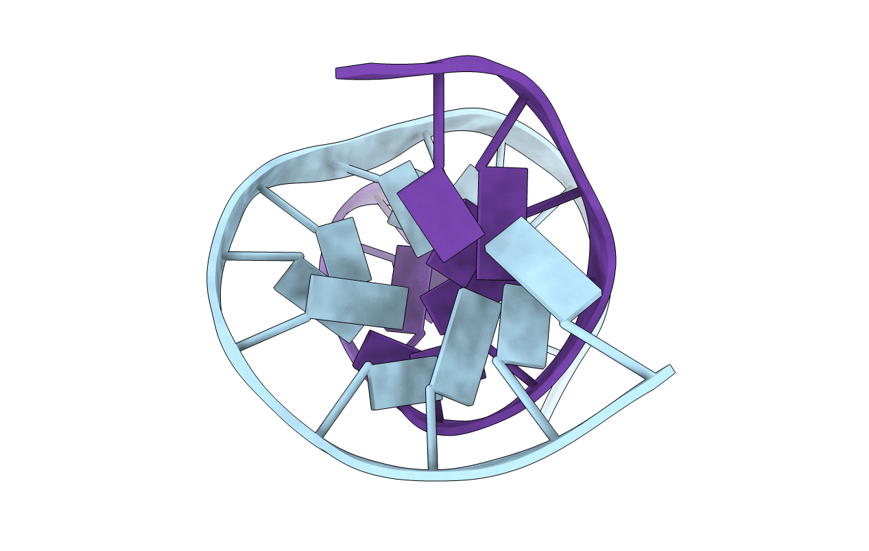
The pyrimidopurinone adduct M1G [3-(2'-deoxy-beta-D-erythro-pentofuranosyl)pyrimido[1,2-a]-purin-10(3H)-one], formed in DNA upon exposure to malondialdehyde or base propenals, was incorporated into 5'-d(ATCGCMCGGCATG)-3'-5'-d(CATGCCGCGAT)-3', where M = M1G. This duplex contained a two-nucleotide bulge in the modified strand, and was named the M1G-2BD oligodeoxynucleotide. It provided a model for -2 bp strand slippage deletions associated with the (CpG)3-iterated repeat hotspot for frameshift mutations from the Salmonella typhimurium hisD3052 gene. M1G was chemically stable in the M1G-2BD duplex at neutral pH. The two-base bulge in the M1G-2BD oligodeoxynucleotide was localized and consisted of M1G and the 3'-neighbor deoxycytosine. The intrahelical orientation of M1G was established from a combination of NOE and chemical shift data. M1G was in the anti conformation about the glycosyl bond. The 3'-neighbor deoxycytosine appeared to be extruded toward the major groove. In contrast, when M1G was placed into the corresponding fully complementary (CpG)3-iterated repeat duplex at neutral pH, spontaneous and quantitative ring-opening to N(2)-(3-oxo-1-propenyl)-dG (the OPG adduct) was facilitated [Mao, H., Reddy, G. R., Marnett, L. J., and Stone, M. P. (1999) Biochemistry 38, 13491-13501]. The structure of the M1G-2BD duplex suggested that the bulged sequence lacked a cytosine amino group properly positioned to facilitate opening of M1G and supports the notion that proper positioning of deoxycytosine complementary to M1G is necessary to promote ring-opening of the exocyclic adduct in duplex DNA. The structure of the M1G-2BD duplex was similar to that of the structural analogue 1,N(2)-propanodeoxyguanosine (PdG) in the corresponding PdG-2BD duplex [Weisenseel, J. P., Moe, J. G., Reddy, G. R., Marnett, L. J., and Stone, M. P. (1995) Biochemistry 34, 50-64]. The fixed position of the bulged bases in both instances suggests that these exocyclic adducts do not facilitate transient bulge migration.