

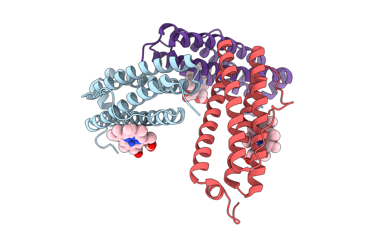
1JGC image
Deposition Date 2001-06-24
Release Date 2002-01-09
Last Version Date 2024-12-25
Entry Detail
PDB ID:
1JGC
Title:
The 2.6 A Structure Resolution of Rhodobacter capsulatus Bacterioferritin with Metal-free Dinuclear Site and Heme Iron in a Crystallographic Special Position
Biological Source:
Source Organism(s):
Method Details:
Experimental Method:
Resolution:
2.60 Å
R-Value Free:
0.24
R-Value Work:
0.22
Space Group:
I 4 2 2

Macromolecular Entities
Structures with similar UniProt ID
Protein Blast
Polymer Type:polypeptide(L)
Molecule:bacterioferritin
Gene (Uniprot):bfr
Chain IDs:A, B, C
Chain Length:161
Number of Molecules:3
Biological Source:Rhodobacter capsulatus
Ligand Molecules
Primary Citation
The 2.6 A resolution structure of Rhodobacter capsulatus bacterioferritin with metal-free dinuclear site and heme iron in a crystallographic 'special position'.
Acta Crystallogr. D Biol. Crystallogr. 58 29 38 (2002)
PMID: 11752777 DOI: 10.1107/S0907444901017267

Abstact

Bacterioferritin from Rhodobacter capsulatus was crystallized and its structure was solved at 2.6 A resolution. This first structure of a bacterioferritin from a photosynthetic organism is a spherical particle of 24 subunits displaying 432 point-group symmetry like ferritin and bacterioferritin from Escherichia coli. Crystallized in the I422 space group, its structural analysis reveals for the first time the non-symmetric heme molecule located on a twofold crystallographic symmetry axis. Other hemes of the protomer are situated on twofold noncrystallographic axes. Apparently, both types of sites bind heme in two orientations, leading to an average structure consisting of a symmetric 50:50 mixture, thus satisfying the crystallographic and noncrystallographic symmetry of the crystal. Five water molecules are situated close to the heme, which is bound in a hydrophobic pocket and axially coordinated by two crystallographic or noncrystallographically related methionine residues. Its ferroxidase center, in which Fe(II) is oxidized to Fe(III), is empty or fractionally occupied by a metal ion. Two positions are observed for the coordinating Glu18 side chain instead of one in the E. coli enzyme in which the site is occupied. This result suggests that the orientation of the Glu18 side chain could be constrained by this interaction.

Legend

Protein

Chemical

Disease

Primary Citation of related structures
Feedback Form
Name
Email
Institute
Feedback