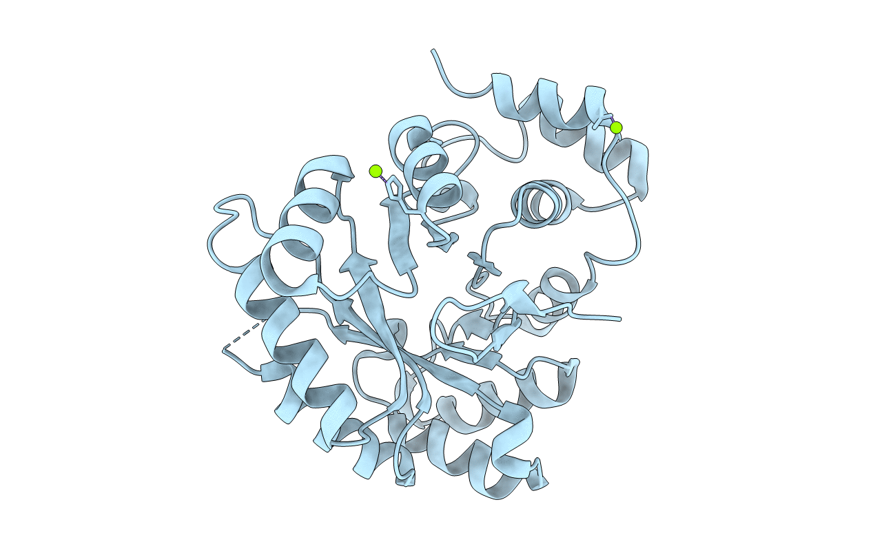
1HW6 image
Deposition Date 2001-01-09
Release Date 2001-06-20
Last Version Date 2023-08-09
Entry Detail
PDB ID:
1HW6
Keywords:
Title:
CRYSTAL STRUCTURE OF APO-2,5-DIKETO-D-GLUCONATE REDUCTASE
Biological Source:
Source Organism(s):
Expression System(s):
Method Details:
Experimental Method:
Resolution:
1.90 Å
Space Group:
P 21 21 21
Macromolecular Entities
Polymer Type:polypeptide(L)
Molecule:2,5-DIKETO-D-GLUCONIC ACID RE
Gene (Uniprot):dkgA
Chain IDs:A
Chain Length:278
Number of Molecules:1
Biological Source:Corynebacterium sp.
Primary Citation
Structural assembly of the active site in an aldo-keto reductase by NADPH cofactor.
J. Mol. Biol. 309 1209 1218 (2001)
PMID: 11399090 DOI: 10.1006/jmbi.2001.4739

Abstact

A 1.9 A resolution X-ray structure of the apo-form of Corynebacterium 2,5-diketo-d-gluconic acid reductase A (2,5-DKGR A), a member of the aldo-keto reductase superfamily, has been determined by molecular replacement using the NADPH-bound form of the same enzyme as the search model. 2,5-DKGR A catalyzes the NADPH-dependent stereo-specific reduction of 2,5-diketo-d-gluconate (2,5-DKG) to 2-keto-l-gulonate, a precursor in the industrial production of vitamin C. An atomic-resolution structure for the apo-form of the enzyme, in conjunction with our previously reported high-resolution X-ray structure for the holo-enzyme and holo/substrate model, allows a comparative analysis of structural changes that accompany cofactor binding. The results show that regions of the active site undergo coordinated conformational changes of up to 8 A. These conformational changes result in the organization and structural rearrangement of residues associated with substrate binding and catalysis. Thus, NADPH functions not only to provide a hydride ion for catalytic reduction, but is also a critical structural component for formation of a catalytically competent form of DKGR A.

Legend

Protein

Chemical

Disease

Primary Citation of related structures
Feedback Form
Name
Email
Institute
Feedback