

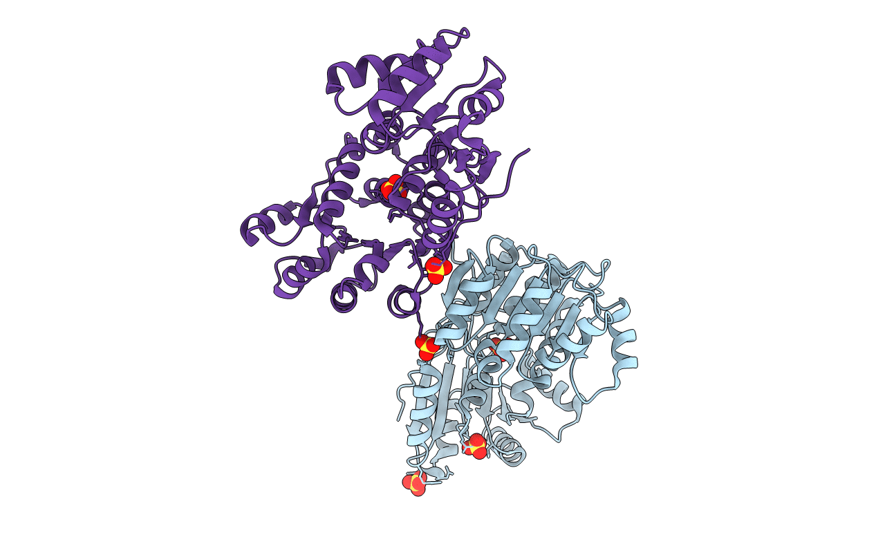
1HV8 image
Deposition Date 2001-01-08
Release Date 2001-02-21
Last Version Date 2024-11-06
Entry Detail
PDB ID:
1HV8
Title:
CRYSTAL STRUCTURE OF A DEAD BOX PROTEIN FROM THE HYPERTHERMOPHILE METHANOCOCCUS JANNASCHII
Biological Source:
Source Organism(s):
Expression System(s):
Method Details:
Experimental Method:
Resolution:
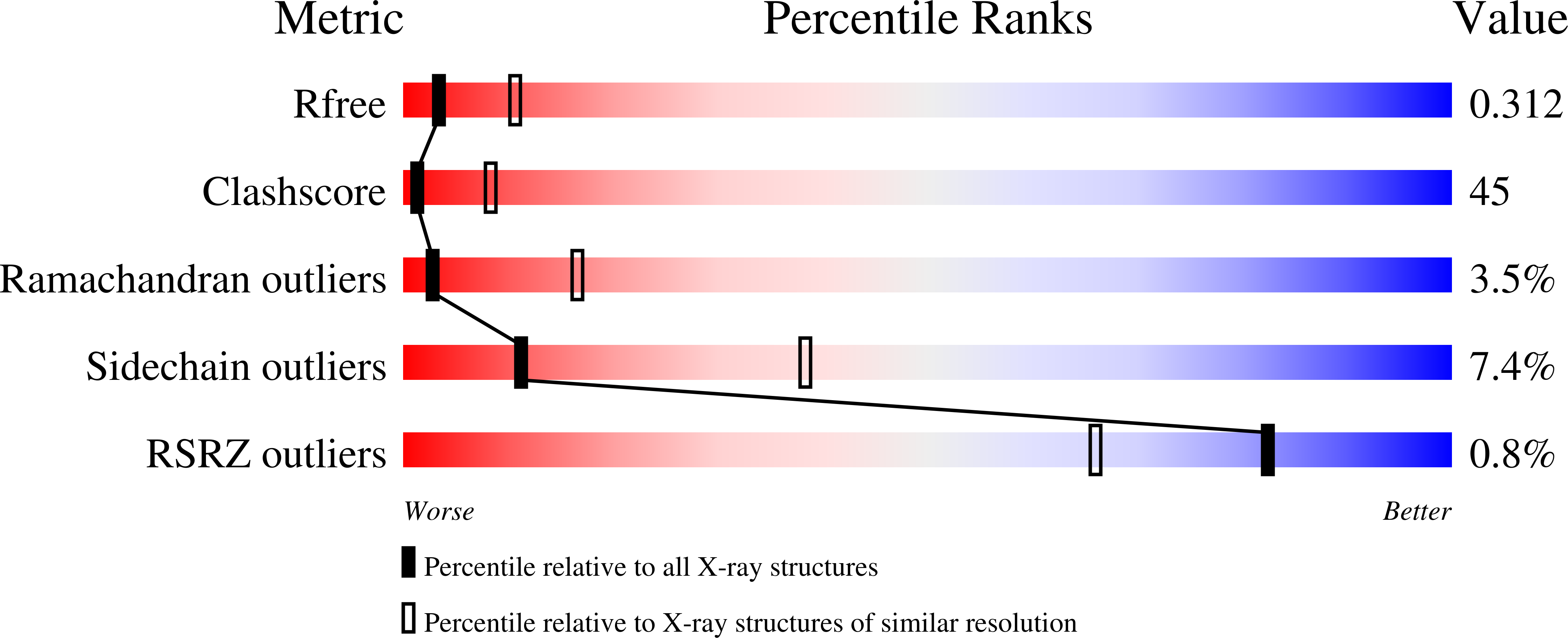
3.00 Å
R-Value Free:
0.31
R-Value Work:
0.27
Space Group:
C 2 2 21
Macromolecular Entities
Structures with similar UniProt ID
Protein Blast
Polymer Type:polypeptide(L)
Molecule:PUTATIVE ATP-DEPENDENT RNA HE
Gene (Uniprot):MJ0669
Chain IDs:A, B
Chain Length:367
Number of Molecules:2
Biological Source:Methanocaldococcus jannaschii
Modified Residue
Compound ID Chain ID Parent Comp ID Details 2D Image
MSE A MET SELENOMETHIONINE
Ligand Molecules
Primary Citation
Crystal structure of a DEAD box protein from the hyperthermophile Methanococcus jannaschii.
Proc. Natl. Acad. Sci. U.S.A. 98 1465 1470 (2001)
PMID: 11171974 DOI: 10.1073/pnas.98.4.1465

Abstact

We have determined the structure of a DEAD box putative RNA helicase from the hyperthermophile Methanococcus jannaschii. Like other helicases, the protein contains two alpha/beta domains, each with a recA-like topology. Unlike other helicases, the protein exists as a dimer in the crystal. Through an interaction that resembles the dimer interface of insulin, the amino-terminal domain's 7-strand beta-sheet is extended to 14 strands across the two molecules. Motifs conserved in the DEAD box family cluster in the cleft between domains, and many of their functions can be deduced by mutational data and by comparison with other helicase structures. Several lines of evidence suggest that motif III Ser-Ala-Thr may be involved in binding RNA.

Legend

Protein

Chemical

Disease

Primary Citation of related structures
Feedback Form
Name
Email
Institute
Feedback