

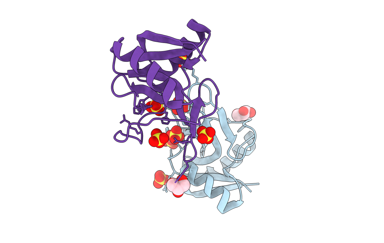
1H8U image
Deposition Date 2001-02-15
Release Date 2001-07-17
Last Version Date 2024-10-23
Entry Detail
PDB ID:
1H8U
Keywords:
Title:
Crystal Structure of the Eosinophil Major Basic Protein at 1.8A: An Atypical Lectin with a Paradigm Shift in Specificity
Biological Source:
Source Organism(s):
HOMO SAPIENS (Taxon ID: 9606)
Method Details:
Experimental Method:
Resolution:
1.80 Å
R-Value Free:
0.26
R-Value Work:
0.23
R-Value Observed:
0.23
Space Group:
C 1 2 1

Macromolecular Entities
Structures with similar UniProt ID
Protein Blast
Polymer Type:polypeptide(L)
Molecule:EOSINOPHIL GRANULE MAJOR BASI
Gene (Uniprot):PRG2
Chain IDs:A, B
Chain Length:117
Number of Molecules:2
Biological Source:HOMO SAPIENS
Primary Citation
Crystal Structure of the Eosinophil Major Basic Protein at 1.8A. An Atypical Lectin with a Paradigm Shift in Specificity
J. Biol. Chem. 276 26197 ? (2001)
PMID: 11319227 DOI: 10.1074/JBC.M100848200

Abstact

The eosinophil major basic protein (EMBP) is the predominant constituent of the crystalline core of the eosinophil primary granule. EMBP is directly implicated in epithelial cell damage, exfoliation, and bronchospasm in allergic diseases such as asthma. Here we report the crystal structure of EMBP at 1.8 A resolution, and show that it is similar to that of members of the C-type lectin superfamily with which it shares minimal amino acid sequence identity (approximately 15--28%). However, this protein lacks a Ca(2+)/carbohydrate-binding site. Our analysis suggests that EMBP specifically binds heparin. Based on our results, we propose a possible new function for this protein, which is likely to have implications for EMBP function.

Legend

Protein

Chemical

Disease

Primary Citation of related structures
Feedback Form
Name
Email
Institute
Feedback