

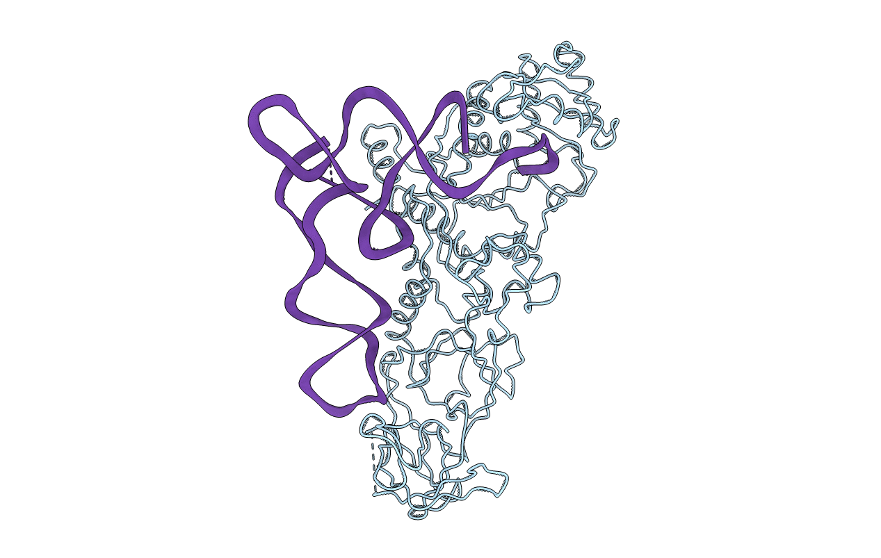
1GSG image
Deposition Date 1990-04-03
Release Date 1992-02-24
Last Version Date 2025-03-19
Entry Detail
PDB ID:
1GSG
Keywords:
Title:
Structure of E.coli glutaminyl-tRNA synthetase complexed with trnagln and ATP at 2.8 Angstroms resolution
Biological Source:
Source Organism(s):
Escherichia coli (Taxon ID: 562)
Method Details:
Experimental Method:
Resolution:
2.80 Å
R-Value Work:
0.27
R-Value Observed:
0.27
Space Group:
C 2 2 21

Macromolecular Entities
Polymer Type:polypeptide(L)
Molecule:GLUTAMINYL-TRNA SYNTHETASE
Gene (Uniprot):glnS
Chain IDs:B (auth: P)
Chain Length:553
Number of Molecules:1
Biological Source:Escherichia coli
Modified Residue
Compound ID Chain ID Parent Comp ID Details 2D Image
2MA A A 2-METHYLADENOSINE-5'-MONOPHOSPHATE
4SU A U 4-THIOURIDINE-5'-MONOPHOSPHATE
5MU A U 5-METHYLURIDINE 5'-MONOPHOSPHATE
H2U A U 5,6-DIHYDROURIDINE-5'-MONOPHOSPHATE
OMG A G O2'-METHYLGUANOSINE-5'-MONOPHOSPHATE
OMU A U O2'-METHYLURIDINE 5'-MONOPHOSPHATE
PSU A U PSEUDOURIDINE-5'-MONOPHOSPHATE
Ligand Molecules
Primary Citation
Structure of E. coli glutaminyl-tRNA synthetase complexed with tRNA(Gln) and ATP at 2.8 A resolution.
Science 246 1135 1142 (1989)
PMID: 2479982

Abstact

The crystal structure of Escherichia coli glutaminyl-tRNA synthetase (GlnRS) complexed with its cognate glutaminyl transfer RNA (tRNA(Gln] and adenosine triphosphate (ATP) has been derived from a 2.8 angstrom resolution electron density map and the known protein and tRNA sequences. The 63.4-kilodalton monomeric enzyme consists of four domains arranged to give an elongated molecule with an axial ratio greater than 3 to 1. Its interactions with the tRNA extend from the anticodon to the acceptor stem along the entire inside of the L of the tRNA. The complexed tRNA retains the overall conformation of the yeast phenylalanine tRNA (tRNA(Phe] with two major differences: the 3' acceptor strand of tRNA(Gln) makes a hairpin turn toward the inside of the L, with the disruption of the final base pair of the acceptor stem, and the anticodon loop adopts a conformation not seen in any of the previously determined tRNA structures. Specific recognition elements identified so far include (i) enzyme contacts with the 2-amino groups of guanine via the tRNA minor groove in the acceptor stem at G2 and G3; (ii) interactions between the enzyme and the anticodon nucleotides; and (iii) the ability of the nucleotides G73 and U1.A72 of the cognate tRNA to assume a conformation stabilized by the protein at a lower free energy cost than noncognate sequences. The central domain of this synthetase binds ATP, glutamine, and the acceptor end of the tRNA as well as making specific interactions with the acceptor stem.2+t is

Legend

Protein

Chemical

Disease

Primary Citation of related structures
Feedback Form
Name
Email
Institute
Feedback