
Deposition Date
1997-01-24
Release Date
1997-08-20
Last Version Date
2024-10-09
Entry Detail
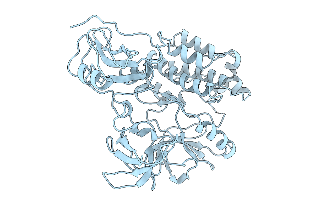
PDB ID:
1FMK
Keywords:
Title:
CRYSTAL STRUCTURE OF HUMAN TYROSINE-PROTEIN KINASE C-SRC
Biological Source:
Source Organism(s):
Homo sapiens (Taxon ID: 9606)
Expression System(s):
Method Details:
Experimental Method:
Resolution:
1.50 Å
R-Value Free:
0.26
R-Value Work:
0.21
R-Value Observed:
0.21
Space Group:
P 21 21 21
