

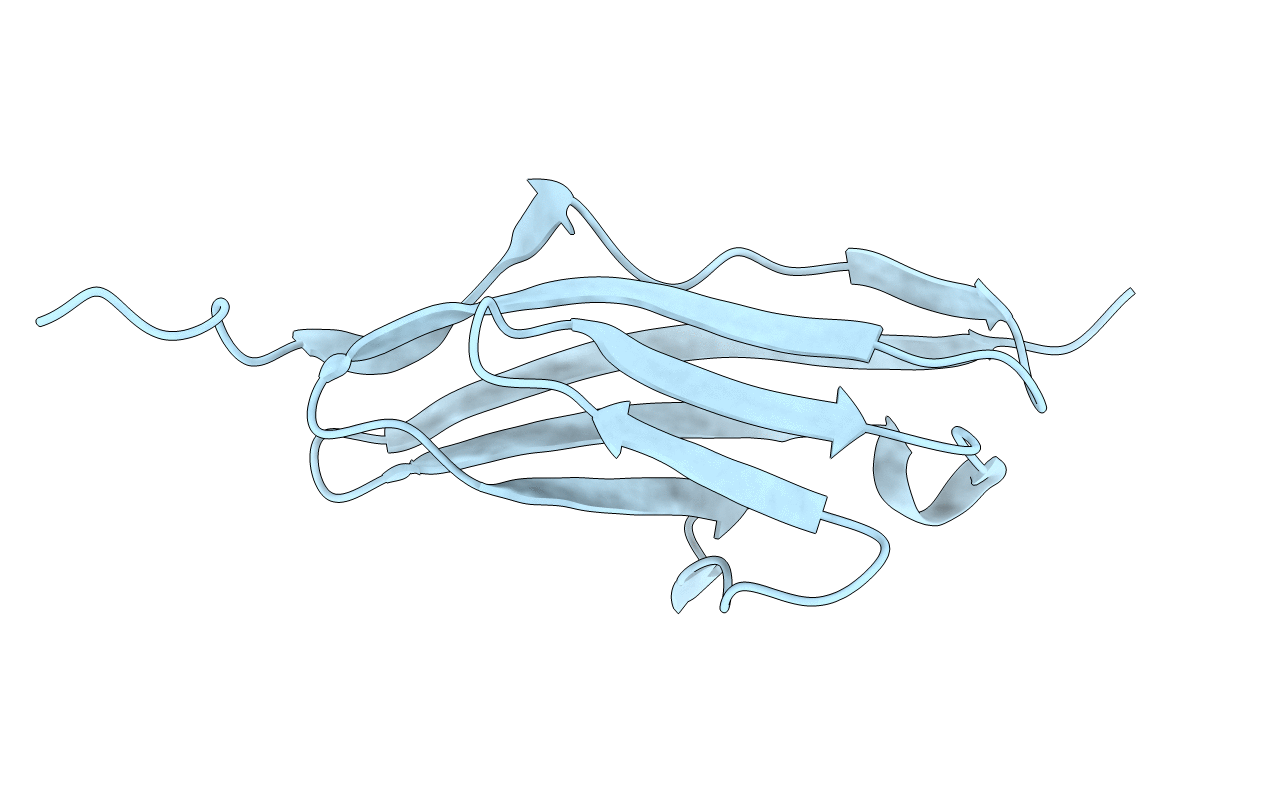
1FHG image
Deposition Date 2000-08-01
Release Date 2000-08-23
Last Version Date 2023-08-09
Entry Detail
PDB ID:
1FHG
Title:
HIGH RESOLUTION REFINEMENT OF TELOKIN
Biological Source:
Source Organism(s):
Method Details:
Experimental Method:
Resolution:
2.00 Å
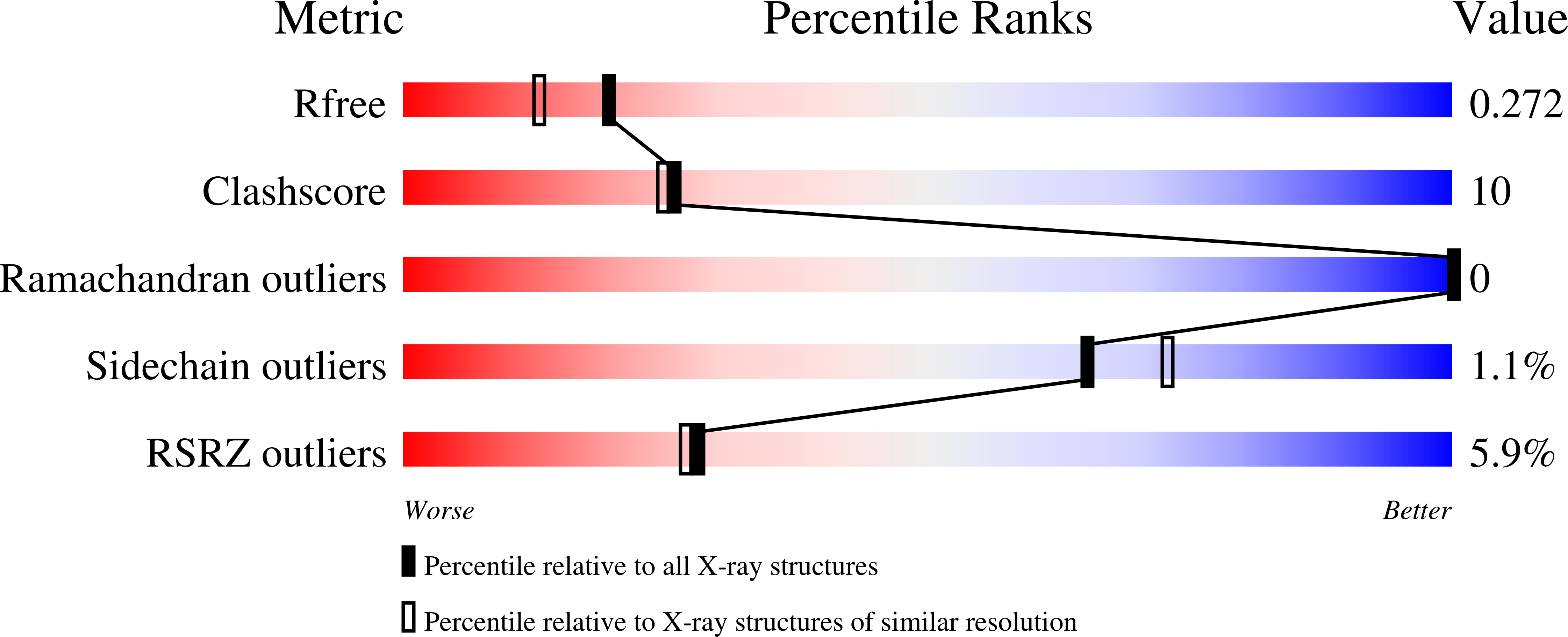
R-Value Free:
0.26
R-Value Work:
0.21
R-Value Observed:
0.23
Space Group:
P 32 2 1
Macromolecular Entities
Polymer Type:polypeptide(L)
Molecule:TELOKIN
Chain IDs:A
Chain Length:154
Number of Molecules:1
Biological Source:Meleagris gallopavo
Primary Citation
X-ray structure determination of telokin, the C-terminal domain of myosin light chain kinase, at 2.8 A resolution.
J. Mol. Biol. 227 840 851 (1992)
PMID: 1404391 DOI: 10.1016/0022-2836(92)90226-A

Abstact

The three-dimensional structure of telokin, an acidic protein identical to the C-terminal portion of smooth muscle myosin light chain kinase from turkey gizzard, has been determined at 2.8 A resolution and refined to a crystallographic R-factor of 19.5% for all measured X-ray data from 30 A to 2.8 A. Crystals used in the investigation belonged to the space group P3(2)21, with one molecule per asymmetric unit and unit cell dimensions of a = b = 64.4 A and c = 50.6 A. Telokin contains 154 amino acid residues, 103 of which were visible in the electron density map. The overall molecular fold of telokin consists of seven strands of antiparallel beta-pleated sheet that wrap around to form a barrel. There is also an extended tail of eight amino acid residues at the N terminus that does not participate in beta-sheet formation. The beta-barrel can be simply envisioned as two layers of beta-sheet, nearly parallel to one another, with one layer containing four and the other three beta-strands. This type of beta-barrel, as seen in telokin, was first observed for the CH2 domain of an immunoglobulin fragment Fc. Telokin is an intracellular protein and, as such, does not contain the disulphide linkage between beta-strands B and F normally observed in the immunoglobulin constant domains. It does, however, contain two cysteine amino acid residues (Cys63 and Cys115) that are situated at structurally identical positions to those forming the disulphide linkage in the immunoglobulin constant domain.

Legend

Protein

Chemical

Disease

Primary Citation of related structures
Feedback Form
Name
Email
Institute
Feedback