1D2Z image
Deposition Date 1999-09-28
Release Date 1999-11-29
Last Version Date 2024-02-07
Entry Detail
PDB ID:
1D2Z
Keywords:
Title:
THREE-DIMENSIONAL STRUCTURE OF A COMPLEX BETWEEN THE DEATH DOMAINS OF PELLE AND TUBE
Biological Source:
Source Organism(s):
Expression System(s):
Method Details:
Experimental Method:
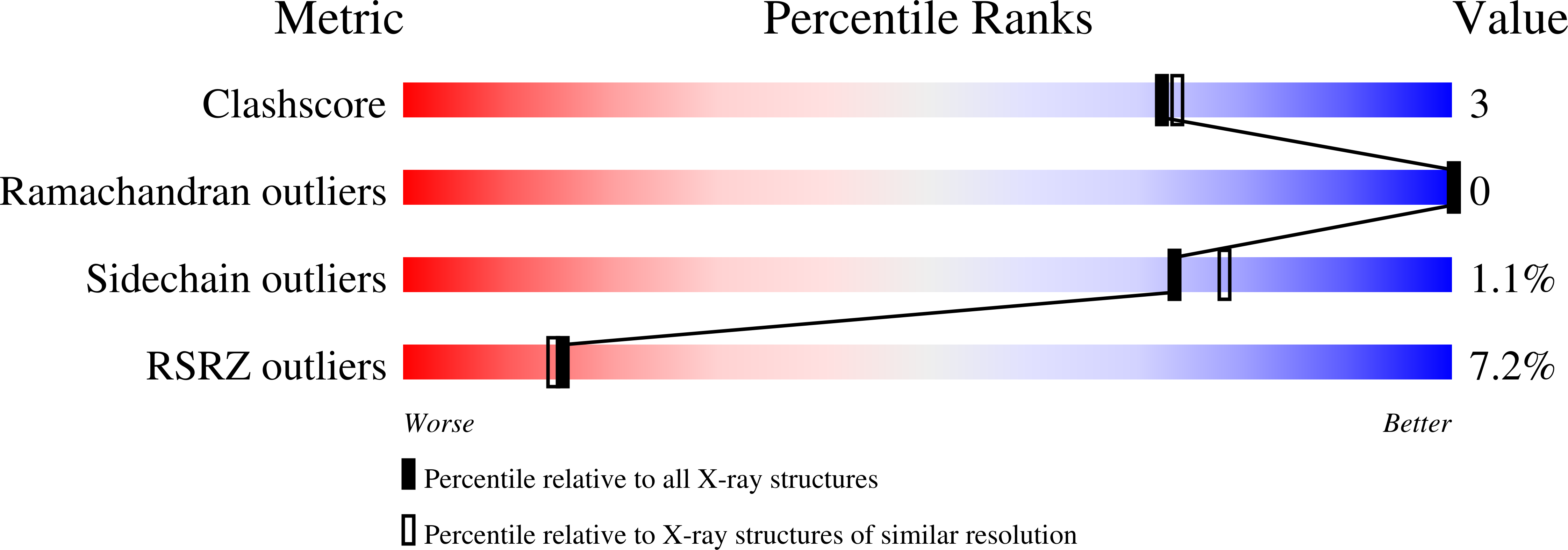
Resolution:
2.00 Å
R-Value Free:
0.24
R-Value Work:
0.21
R-Value Observed:
0.22
Space Group:
P 21 21 21
Macromolecular Entities
Structures with similar UniProt ID
Protein Blast
Polymer Type:polypeptide(L)
Molecule:DEATH DOMAIN OF PELLE
Gene (Uniprot):pll
Chain IDs:A, C
Chain Length:108
Number of Molecules:2
Biological Source:Drosophila melanogaster
Structures with similar UniProt ID
Protein Blast
Polymer Type:polypeptide(L)
Molecule:DEATH DOMAIN OF TUBE
Gene (Uniprot):tub
Chain IDs:B, D
Chain Length:153
Number of Molecules:2
Biological Source:Drosophila melanogaster
Ligand Molecules
Primary Citation
Three-dimensional structure of a complex between the death domains of Pelle and Tube.
Cell 99 545 555 (1999)
PMID: 10589682 DOI: 10.1016/S0092-8674(00)81542-1

Abstact

The interaction of the serine/threonine kinase Pelle and adaptor protein Tube through their N-terminal death domains leads to the nuclear translocation of the transcription factor Dorsal and activation of zygotic patterning genes during Drosophila embryogenesis. Crystal structure of the Pelle and Tube death domain heterodimer reveals that the two death domains adopt a six-helix bundle fold and are arranged in an open-ended linear array with plastic interfaces mediating their interactions. The Tube death domain has an insertion between helices 2 and 3, and a C-terminal tail making significant and indispensable contacts in the heterodimer. In vivo assays of Pelle and Tube mutants confirmed that the integrity of the major heterodimer interface is critical to the activity of these molecules.

Legend

Protein

Chemical

Disease

Primary Citation of related structures
Feedback Form
Name
Email
Institute
Feedback