

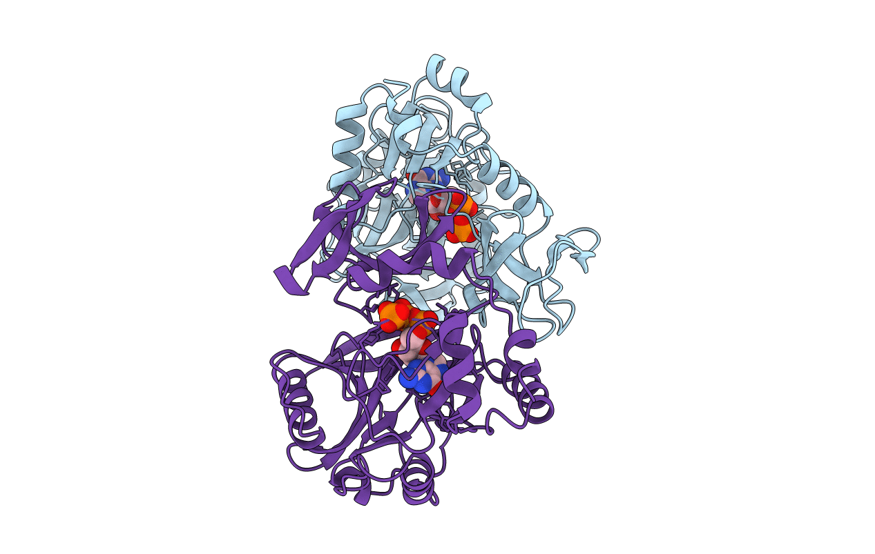
1CKM image
Deposition Date 1997-04-20
Release Date 1997-07-07
Last Version Date 2024-02-07
Entry Detail
PDB ID:
1CKM
Keywords:
Title:
STRUCTURE OF TWO DIFFERENT CONFORMATIONS OF MRNA CAPPING ENZYME IN COMPLEX WITH GTP
Biological Source:
Source Organism(s):
Expression System(s):
Method Details:
Experimental Method:
Resolution:
2.50 Å
R-Value Free:
0.29
R-Value Work:
0.21
R-Value Observed:
0.21
Space Group:
C 2 2 21
Macromolecular Entities
Structures with similar UniProt ID
Protein Blast
Polymer Type:polypeptide(L)
Molecule:MRNA CAPPING ENZYME
Gene (Uniprot):A103R
Chain IDs:A, B
Chain Length:330
Number of Molecules:2
Biological Source:Paramecium bursaria Chlorella virus 1
Ligand Molecules
Primary Citation
X-ray crystallography reveals a large conformational change during guanyl transfer by mRNA capping enzymes.
Cell 89 545 553 (1997)
PMID: 9160746 DOI: 10.1016/S0092-8674(00)80236-6

Abstact

We have solved the crystal structure of an mRNA capping enzyme at 2.5 A resolution. The enzyme comprises two domains with a deep, but narrow, cleft between them. The two molecules in the crystallographic asymmetric unit adopt very different conformations; both contain a bound GTP, but one protein molecule is in an open conformation while the other is in a closed conformation. Only in the closed conformation is the enzyme able to bind manganese ions and undergo catalysis within the crystals to yield the covalent guanylated enzyme intermediate. These structures provide direct evidence for a mechanism that involves a significant conformational change in the enzyme during catalysis.

Legend

Protein

Chemical

Disease

Primary Citation of related structures
Feedback Form
Name
Email
Institute
Feedback