

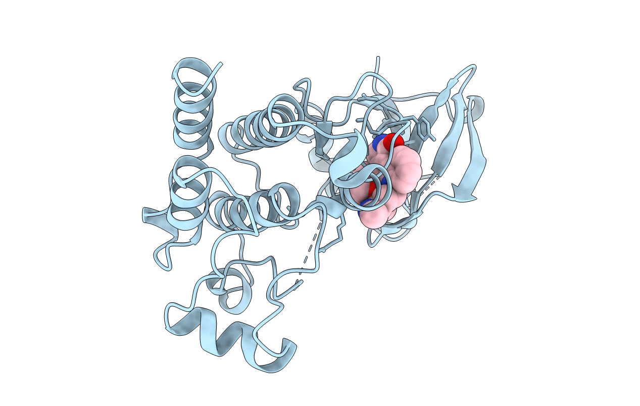
1BYG image
Deposition Date 1998-10-14
Release Date 1999-10-14
Last Version Date 2023-08-09
Entry Detail
PDB ID:
1BYG
Keywords:
Title:
KINASE DOMAIN OF HUMAN C-TERMINAL SRC KINASE (CSK) IN COMPLEX WITH INHIBITOR STAUROSPORINE
Biological Source:
Source Organism(s):
Homo sapiens (Taxon ID: 9606)
Expression System(s):
Method Details:
Experimental Method:
Resolution:
2.40 Å
R-Value Free:
0.28
R-Value Work:
0.19
Space Group:
P 21 21 2
Macromolecular Entities
Polymer Type:polypeptide(L)
Molecule:PROTEIN (C-TERMINAL SRC KINAS
Gene (Uniprot):CSK
Chain IDs:A
Chain Length:278
Number of Molecules:1
Biological Source:Homo sapiens
Ligand Molecules
Primary Citation
Structure of the protein tyrosine kinase domain of C-terminal Src kinase (CSK) in complex with staurosporine.
J. Mol. Biol. 285 713 725 (1999)
PMID: 9878439 DOI: 10.1006/jmbi.1998.2369

Abstact

The crystal structure of the kinase domain of C-terminal Src kinase (CSK) has been determined by molecular replacement, co-complexed with the protein kinase inhibitor staurosporine (crystals belong to the space group P21212 with a=44.5 A, b=120.6 A, c=48.3 A). The final model of CSK has been refined to an R-factor of 19.9 % (Rfree=28.7 %) at 2.4 A resolution. The structure consists of a small, N-terminal lobe made up mostly of a beta-sheet, and a larger C-terminal lobe made up mostly of alpha-helices. The structure reveals atomic details of interactions with staurosporine, which binds in a deep cleft between the lobes. The polypeptide chain fold of CSK is most similar to c-Src, Hck and fibroblast growth factor receptor 1 kinase (FGFR1K) and most dissimilar to insulin receptor kinase (IRK). Interactions between the N and C-terminal lobe are mediated by the bound staurosporine molecule and by hydrogen bonds. In addition, there are several water molecules forming lobe-bridging hydrogen bonds, which may be important for maintaining the catalytic integrity of the kinase. Furthermore, the conserved Lys328 and Glu267 residues utilise water in the formation of a molecular pivot which is essential in allowing relative movement of the N and C-terminal lobes. An analysis of the residues around the ATP-binding site reveals structural differences with other protein tyrosine kinases. Most notable of these are different orientations of the conserved residues Asp332 and Phe333, suggesting that inhibitor binding proceeds via an induced fit. These structural observations have implications for understanding protein tyrosine kinase catalytic mechanisms and for the design of ATP-mimicking inhibitors of protein kinases.

Legend

Protein

Chemical

Disease

Primary Citation of related structures
Feedback Form
Name
Email
Institute
Feedback