

1BQU image
Deposition Date 1998-08-18
Release Date 1998-08-26
Last Version Date 2024-11-20
Entry Detail
PDB ID:
1BQU
Title:
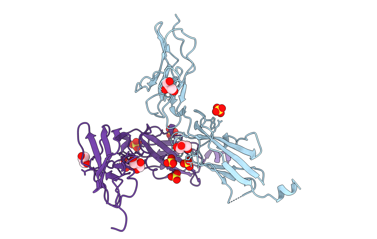
CYTOKYNE-BINDING REGION OF GP130
Biological Source:
Source Organism(s):
Homo sapiens (Taxon ID: 9606)
Expression System(s):
Method Details:
Experimental Method:
Resolution:
2.00 Å
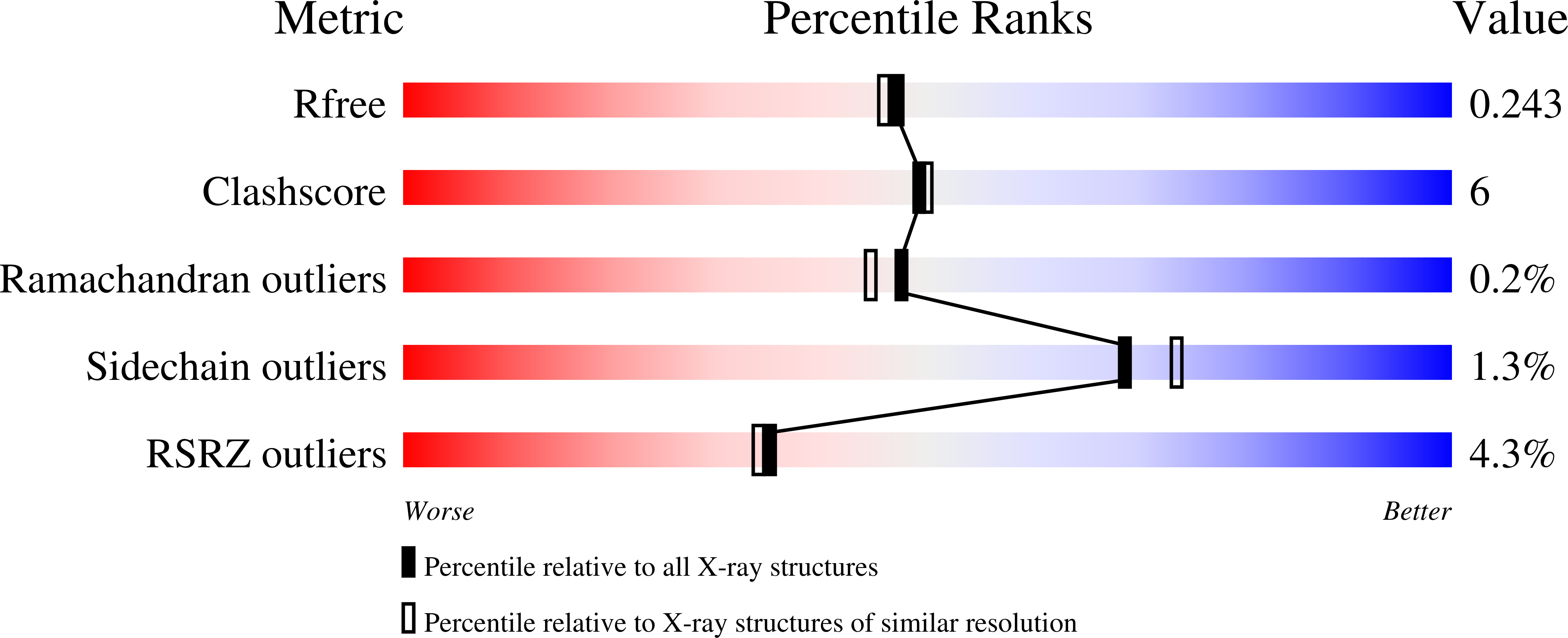
R-Value Free:
0.25
R-Value Work:
0.22
R-Value Observed:
0.22
Space Group:
C 2 2 21
Macromolecular Entities
Structures with similar UniProt ID
Protein Blast
Polymer Type:polypeptide(L)
Molecule:PROTEIN (GP130)
Gene (Uniprot):IL6ST
Chain IDs:A, B
Chain Length:215
Number of Molecules:2
Biological Source:Homo sapiens
Primary Citation
Crystal structure of a cytokine-binding region of gp130.
EMBO J. 17 1665 1674 (1998)
PMID: 9501088 DOI: 10.1093/emboj/17.6.1665

Abstact

The structure of the cytokine-binding homology region of the cell surface receptor gp130 has been determined by X-ray crystallography at 2.0 A resolution. The beta sandwich structure of the two domains conforms to the topology of the cytokine receptor superfamily. This first structure of an uncomplexed receptor exhibits a similar L-shaped quaternary structure to that of ligand-bound family members and suggests a limited flexibility in relative domain orientation of some 3 degrees. The putative ligand-binding loops are relatively rigid, with a phenylalanine side chain similarly positioned to exposed aromatic residues implicated in ligand binding for other such receptors. The positioning and structure of the N-terminal portion of the polypeptide chain have implications for the structure and function of cytokine receptors, such as gp130, which contain an additional N-terminal immunoglobulin-like domain.

Legend

Protein

Chemical

Disease

Primary Citation of related structures
Feedback Form
Name
Email
Institute
Feedback