Deposition Date
1994-12-12
Release Date
1996-04-04
Last Version Date
2024-10-30
Entry Detail
PDB ID:
1BND
Keywords:
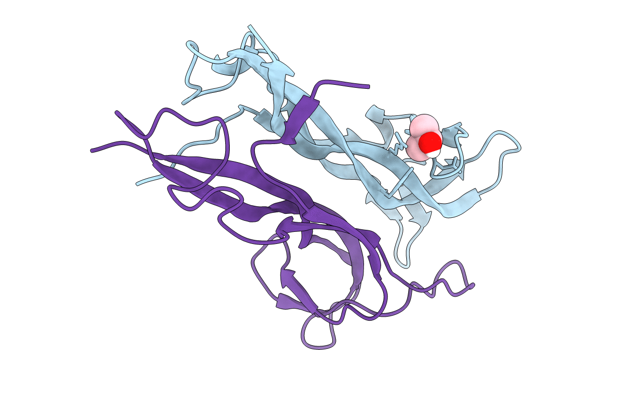
Title:
STRUCTURE OF THE BRAIN-DERIVED NEUROTROPHIC FACTOR(SLASH)NEUROTROPHIN 3 HETERODIMER
Biological Source:
Source Organism(s):
Homo sapiens (Taxon ID: 9606)
Expression System(s):
Method Details:
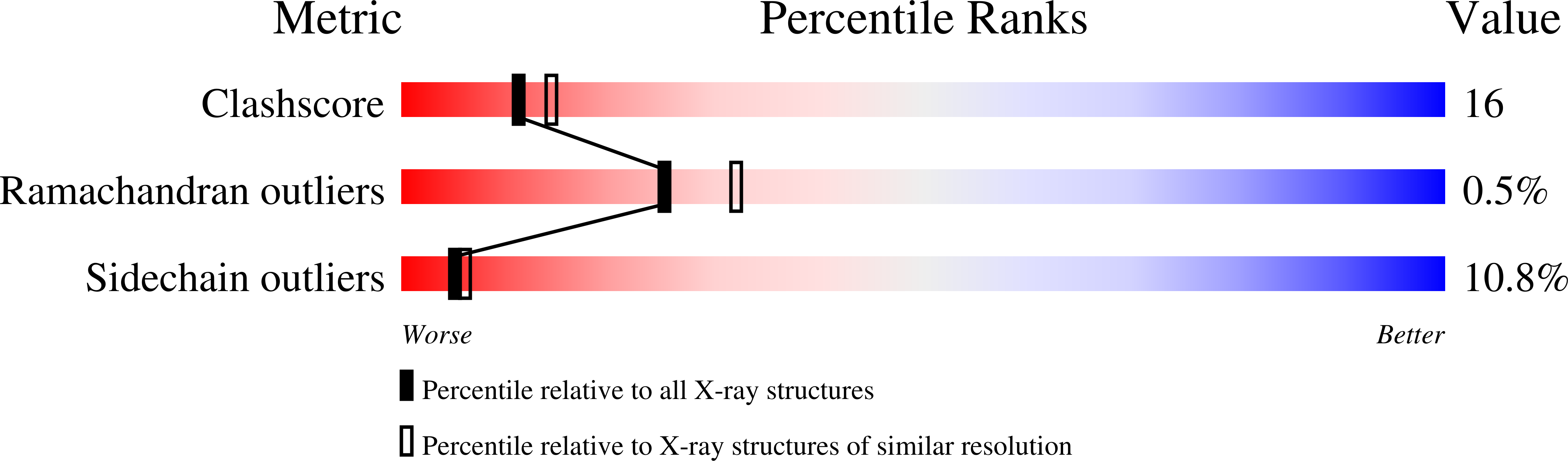
Experimental Method:
Resolution:
2.30 Å
R-Value Work:
0.18
R-Value Observed:
0.18
Space Group:
C 1 2 1