

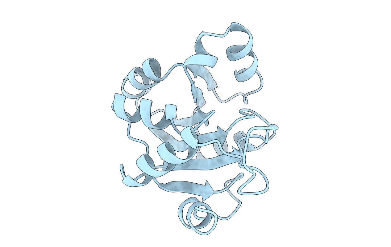
1BLE image
Deposition Date 1997-05-15
Release Date 1997-09-17
Last Version Date 2024-02-07
Entry Detail
PDB ID:
1BLE
Title:
PHOSPHOENOLPYRUVATE-DEPENDENT PHOSPHOTRANSFERASE SYSTEM
Biological Source:
Source Organism(s):
Expression System(s):
Method Details:
Experimental Method:
Resolution:
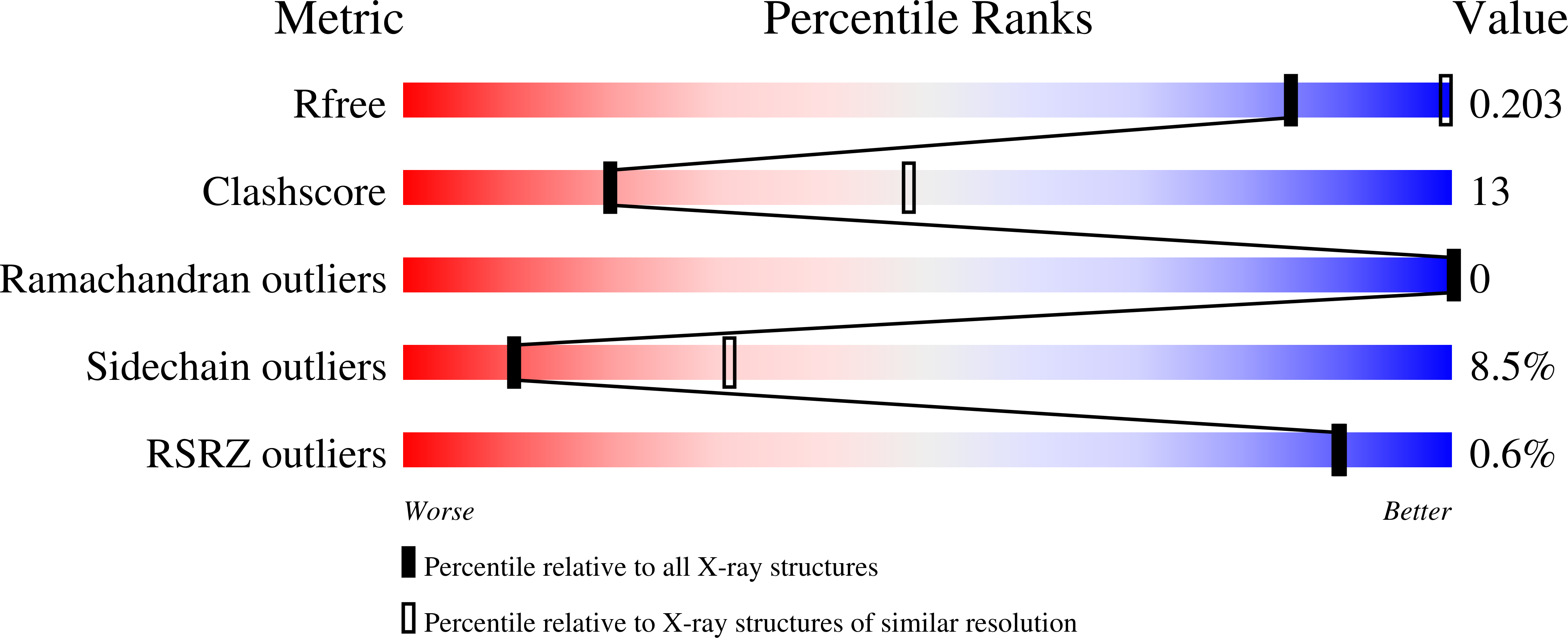
2.90 Å
R-Value Free:
0.26
R-Value Work:
0.18
R-Value Observed:
0.18
Space Group:
P 43 3 2
Macromolecular Entities
Structures with similar UniProt ID
Protein Blast
Polymer Type:polypeptide(L)
Molecule:FRUCTOSE PERMEASE
Gene (Uniprot):levE
Chain IDs:A
Chain Length:163
Number of Molecules:1
Biological Source:Bacillus subtilis
Primary Citation
Crystal structure of the IIB subunit of a fructose permease (IIBLev) from Bacillus subtilis.
J. Mol. Biol. 276 591 602 (1998)
PMID: 9551099 DOI: 10.1006/jmbi.1997.1544

Abstact

The bacterial phosphoenolpyruvate-dependent phosphotransferase system (PTS) mediates both the uptake of carbohydrates across the cytoplasmic membrane and their phosphorylation. During this process, a phosphoryl group is transferred from phosphoenolpyruvate via the general PTS proteins enzyme I, HPr and the sugar-specific components IIA, IIB to the transported sugar. The crystal structure of the IIB subunit of a fructose transporter from Bacillus subtilis (IIBLev) was solved by MIRAS to a resolution of 2.9 A. IIBLev comprises 163 amino acid residues that are folded into an open, mainly parallel beta-sheet with helices packed on either face. The phosphorylation site (His15) is located on the first loop (1/A) at one of the topological switch-points of the fold. Despite different global folds, IIBLev and HPr have very similar active-site loop conformations with the active-site histidine residues located close to the N terminus of the first helix. This resemblance may be of functional importance, since both proteins exchange a phosphoryl group with the same IIA subunit. The structural basis of phosphoryl transfer from HPr to IIAMan to IIBMan was investigated by modeling of the respective transition state complexes using the known HPr and IIAMan structures and a homology model of IIBMan that was derived from the IIBLev structure. All three proteins contain a helix that appears to be suitable for stabilization of the phospho-histidine by dipole and H-bonding interactions. Smooth phosphoryl transfer from one N-cap position to the other appears feasible with a minimized transition state energy due to simultaneous interactions with the donor and the acceptor helix.

Legend

Protein

Chemical

Disease

Primary Citation of related structures
Feedback Form
Name
Email
Institute
Feedback