Deposition Date
1997-10-02
Release Date
1998-10-28
Last Version Date
2024-02-07
Entry Detail
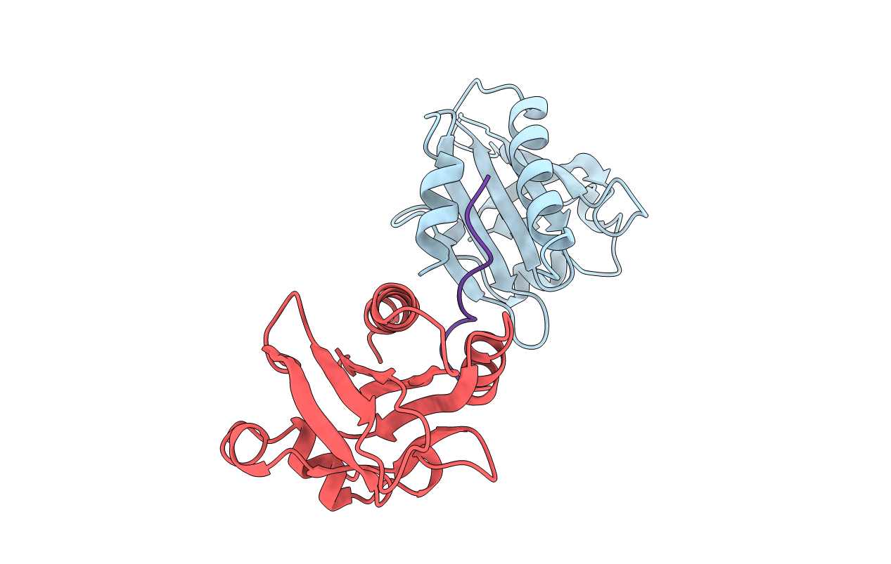
PDB ID:
1AWI
Keywords:
Title:
HUMAN PLATELET PROFILIN COMPLEXED WITH THE L-PRO10 PEPTIDE
Biological Source:
Source Organism(s):
Homo sapiens (Taxon ID: 9606)
Expression System(s):
Method Details:
Experimental Method:
Resolution:
2.20 Å
R-Value Free:
0.30
R-Value Work:
0.20
R-Value Observed:
0.20
Space Group:
P 21 21 2