

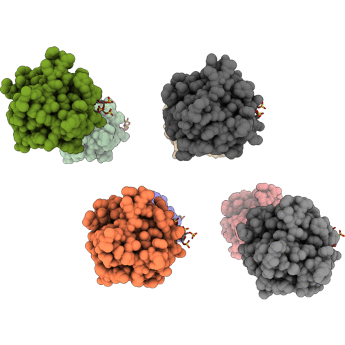
1AFC image
Deposition Date 1993-07-13
Release Date 1993-10-31
Last Version Date 2024-02-07
Entry Detail
PDB ID:
1AFC
Keywords:
Title:
STRUCTURAL STUDIES OF THE BINDING OF THE ANTI-ULCER DRUG SUCROSE OCTASULFATE TO ACIDIC FIBROBLAST GROWTH FACTOR
Biological Source:
Source Organism(s):
Bos taurus (Taxon ID: 9913)
Expression System(s):
Method Details:
Experimental Method:
Resolution:
2.70 Å
R-Value Work:
0.20
R-Value Observed:
0.20
Space Group:
P 32 2 1

Macromolecular Entities
Polymer Type:polypeptide(L)
Molecule:ACIDIC FIBROBLAST GROWTH FACT
Gene (Uniprot):FGF1
Chain IDs:A, B, C, D, E, F, G, H
Chain Length:140
Number of Molecules:8
Biological Source:Bos taurus
Ligand Molecules
Peptide-like Molecules
PRD_900013
Primary Citation
Structural studies of the binding of the anti-ulcer drug sucrose octasulfate to acidic fibroblast growth factor.
Structure 1 27 34 (1993)
PMID: 7520817 DOI: 10.1016/0969-2126(93)90006-3

Abstact

BACKGROUND The anti-ulcer drug sucrose octasulfate (SOS) binds to fibroblast growth factors (FGFs), proteins which stimulate the growth and differentiation of several cell types, including stomach epithelial cells. It is believed that SOS stabilizes FGFs against acid denaturation in the stomach, thus enhancing their ability to stimulate healing of ulcerated tissue. SOS binds to the same site on FGF as heparin and other proteoglycans; in vivo, FGF must bind to cell-surface proteoglycans or to heparin before it can interact with FGF receptors and stimulate growth. The details of this process are not understood. RESULTS We report the crystal structure of a 1:1 complex between acidic FGF (aFGF) and SOS at 2.7 A resolution. SOS binds to a positively charged region of aFGF, largely composed of residues 112-127, and makes contacts primarily with Lys112, Arg116, Lys118, and Arg122. This region is also important in binding heparin. The overall conformation of aFGF is not changed by binding SOS, although the positions of some side chains in the binding site shift by as much as 6 A. CONCLUSION The SOS-FGF crystal structure is consistent with the model that SOS stabilizes FGF by neutralizing several positively charged residues that would destabilize the native structure by electrostatic repulsion. On the basis of this structure, we provide a model for the complex of heparin with an FGF dimer. Such interactions may facilitate FGF receptor dimerization, which may be important in receptor signaling.

Legend

Protein

Chemical

Disease

Primary Citation of related structures
Feedback Form
Name
Email
Institute
Feedback