

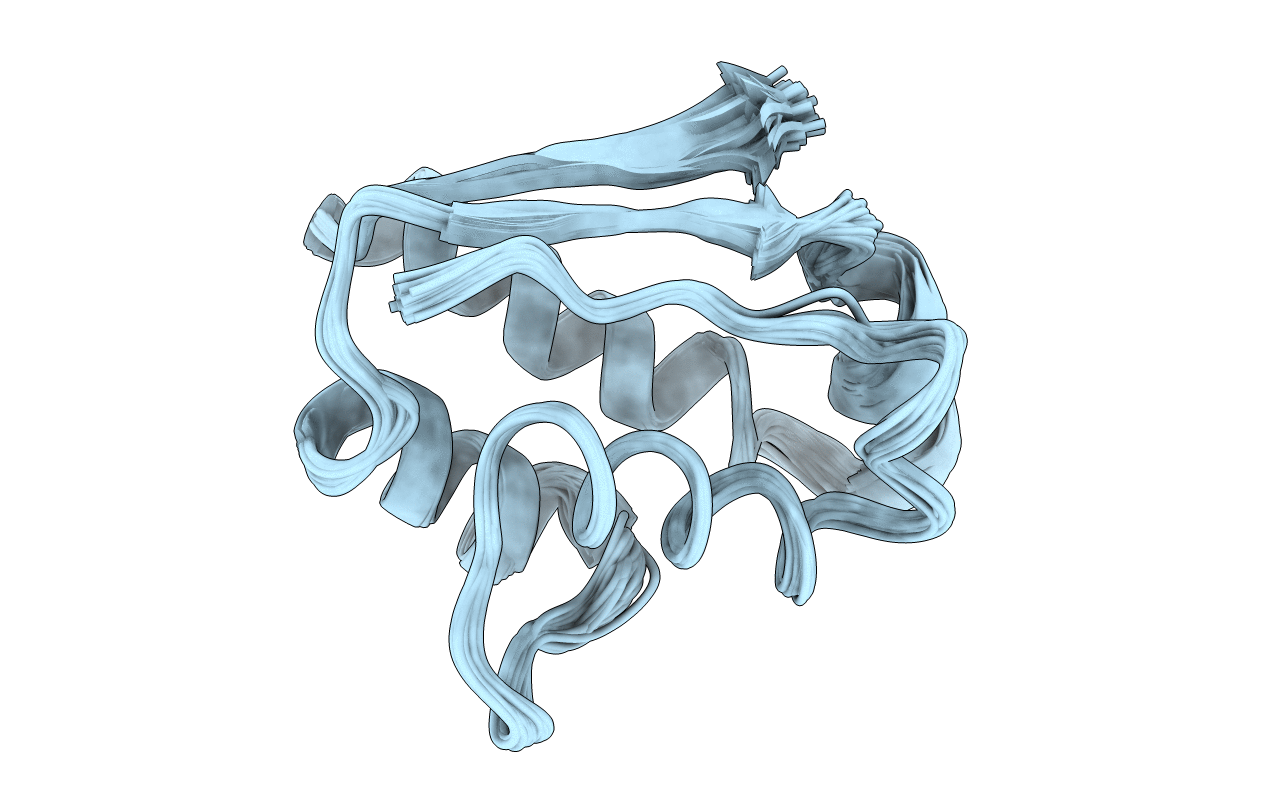
1AB7 image
Deposition Date 1997-02-04
Release Date 1997-09-04
Last Version Date 2024-05-22
Entry Detail
PDB ID:
1AB7
Title:
NMR 15N RELAXATION AND STRUCTURAL STUDIES REVEAL CONFORMATIONAL EXCHANGE IN BARSTAR C40/82A, 30 STRUCTURES
Biological Source:
Source Organism(s):
Expression System(s):
Method Details:
Experimental Method:
Conformers Calculated:
150
Conformers Submitted:
30
Selection Criteria:
LEAST RESTRAINT VIOLATION

Macromolecular Entities
Structures with similar UniProt ID
Protein Blast
Polymer Type:polypeptide(L)
Molecule:BARSTAR
Mutagens:C40A, C82A
Chain IDs:A
Chain Length:89
Number of Molecules:1
Biological Source:Bacillus amyloliquefaciens
Ligand Molecules
Primary Citation
NMR 15N relaxation and structural studies reveal slow conformational exchange in barstar C40/82A.
J. Mol. Biol. 268 494 511 (1997)
PMID: 9159486 DOI: 10.1006/jmbi.1997.0989

Abstact

Barstar an 89-residue protein consisting of four helices and a three-stranded parallel beta-sheet, is the intracellular inhibitor of the endoribonuclease barnase. Barstar C40/82A, a mutant in which the two cysteine residues have been replaced by alanine, has been used as a pseudo wild-type in folding studies and in the crystal structure of the barnase:barstar C40/82A complex. We have determined a high resolution solution structure of barstar C40/82A. The structures of barstar C40/82A and the wild-type are superimposable. A comparison with the crystal structure of the barnase:barstar C40/82A complex revealed subtle differences in the regions involved in the binding of barstar to barnase. Side-chain rotations of residues Asn33, Asp35 and Asp39 and a movement of the binding loop (Pro27-Glu32) towards the binding site of barnase facilitate the formation of interface hydrogen bonds and aromatic contacts in the complex. Extreme line broadening and missing signals in 1H-15N correlation spectra indicate substantial conformational exchange for a large subset of residues. 15N relaxation data at two magnetic field strengths, 11.74 T and 14.10 T, were used to estimate exchange contributions and to map the spectral density function at five frequencies: 0, 50, 60, 450 and 540 MHz. Based on these results, model-free calculations with the inclusion of estimated exchange contributions were used to derive order parameters and internal correlation times. The validity of this approach has been investigated with model-free calculations that incorporate longitudinal relaxation rates and heteronuclear 1H-15N NOE data only at 11.74 T and 14.10 T. The relaxation data suggest substantial conformational exchange in regions of barstar C40/82A, including the binding loop, the second and the third helices, and the second and the third strands. Amide proton exchange experiments suggest a stable hydrogen bond network for all helices and sheets except the third helix and the C-terminal of the second and the third strands. The combined results indicate a rigid body movement of the second helix and twisting motions of the beta-sheet of barstar, which might be important for the interaction with barnase.

Legend

Protein

Chemical

Disease

Primary Citation of related structures
Feedback Form
Name
Email
Institute
Feedback