

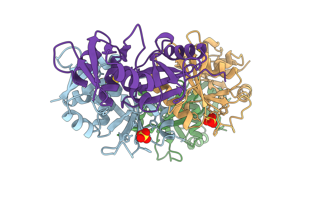
1A79 image
Deposition Date 1998-03-23
Release Date 1999-06-01
Last Version Date 2024-02-07
Entry Detail
PDB ID:
1A79
Keywords:
Title:
CRYSTAL STRUCTURE OF THE TRNA SPLICING ENDONUCLEASE FROM METHANOCOCCUS JANNASCHII
Biological Source:
Source Organism(s):
Expression System(s):
Method Details:
Experimental Method:
Resolution:
2.28 Å
R-Value Free:
0.26
R-Value Work:
0.20
R-Value Observed:
0.20
Space Group:
P 21 21 21

Macromolecular Entities
Structures with similar UniProt ID
Protein Blast
Polymer Type:polypeptide(L)
Molecule:TRNA ENDONUCLEASE
Gene (Uniprot):endA
Chain IDs:A, B, C, D
Chain Length:171
Number of Molecules:4
Biological Source:Methanocaldococcus jannaschii
Primary Citation
Crystal structure and evolution of a transfer RNA splicing enzyme.
Science 280 279 284 (1998)
PMID: 9535656 DOI: 10.1126/science.280.5361.279

Abstact

The splicing of transfer RNA precursors is similar in Eucarya and Archaea. In both kingdoms an endonuclease recognizes the splice sites and releases the intron, but the mechanism of splice site recognition is different in each kingdom. The crystal structure of the endonuclease from the archaeon Methanococcus jannaschii was determined to a resolution of 2.3 angstroms. The structure indicates that the cleavage reaction is similar to that of ribonuclease A and the arrangement of the active sites is conserved between the archaeal and eucaryal enzymes. These results suggest an evolutionary pathway for splice site recognition.

Legend

Protein

Chemical

Disease

Primary Citation of related structures
Feedback Form
Name
Email
Institute
Feedback