

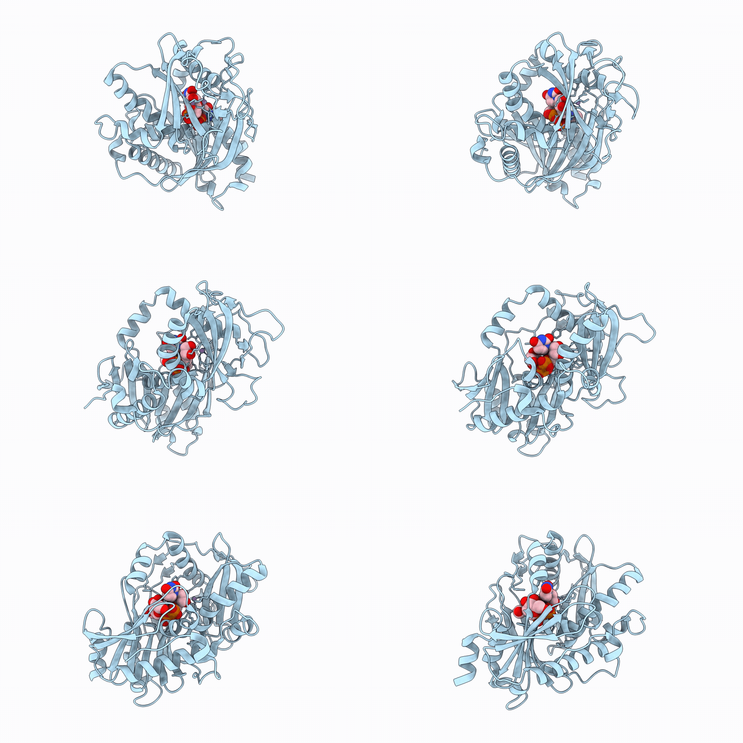
11AP image
Deposition Date 2026-02-13
Release Date 2026-06-10
Last Version Date 2026-06-10
Entry Detail
PDB ID:
11AP
Title:
Crystal structure of mouse DXO in complex with the UDP-N-acetylglucosamine cap
Biological Source:
Source Organism(s):
Mus musculus (Taxon ID: 10090)
Expression System(s):
Method Details:
Experimental Method:
Resolution:
1.80 Å
R-Value Free:
0.20
R-Value Work:
0.16
R-Value Observed:
0.16
Space Group:
P 1 21 1

Macromolecular Entities
Polymer Type:polypeptide(L)
Molecule:Decapping and exoribonuclease
Gene (Uniprot):Dxo
Chain IDs:A
Chain Length:358
Number of Molecules:1
Biological Source:Mus musculus
Primary Citation
Crystal structure of mouse DXO in complex with the UDP-N-acetylglucosamine cap and molecular mechanism for the decapping reactions.
Nucleic Acids Res. 54 ? ? (2026)
PMID: 42200294 DOI: 10.1093/nar/gkag521

Abstact

Noncanonical metabolite 5' caps have recently been identified on RNAs, and the DXO/Rai1 family of enzymes can remove these caps in eukaryotes. While the binding modes of NAD, FAD and dephospho-CoA (dpCoA) caps in the active site of mouse DXO have been determined, how DXO recognizes the UDP-glucose (UDP-Glc) and UDP-N-acetylglucosamine (UDP-GlcNAc) caps is not known. In addition, the molecular mechanism by which DXO catalyzes the decapping reactions is still poorly understood, especially the location of the water/hydroxide that attacks the scissile phosphate to initiate the decapping. Here we report the crystal structure of mouse DXO in complex with UDP-GlcNAc at 1.8 A resolution. The binding mode of the compound explains why DXO removes the entire cap from RNA. We have also determined the structures of mouse DXO in complex with purine oligonucleotides, pA5 and pGGGUU. Most importantly, we have produced a model of DXO in a catalytically competent complex with substrates, revealing that a water/hydroxide coordinated to the first metal ion is the nucleophile that attacks the scissile phosphate. The conformation of the scissile phosphate is similar to an alternate conformer of the 5' phosphate in pA5, which provides experimental support for the modeled substrate complex.

Legend

Protein

Chemical

Disease

Primary Citation of related structures
Feedback Form
Name
Email
Institute
Feedback