

Search Count: 16
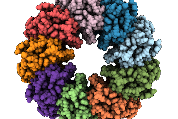 |
Netf 9Mer Pre-Pore Structure On 2N2 Nanodiscs.
Organism: Clostridium perfringens
Method: ELECTRON MICROSCOPY Resolution:2.13 Å Release Date: 2026-02-18 Classification: TOXIN |
Organism: Clostridium perfringens
Method: ELECTRON MICROSCOPY
Release Date: 2026-02-18
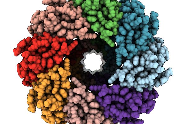 |
Netf Pore Structure
Organism: Clostridium perfringens
Method: ELECTRON MICROSCOPY Resolution:2.10 Å Release Date: 2026-02-18 Classification: TOXIN |
Organism: Clostridium perfringens
Method: ELECTRON MICROSCOPY
Release Date: 2026-02-18
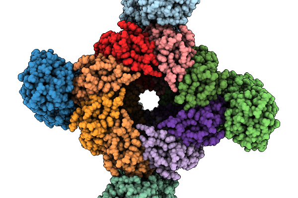 |
Netf - Antxr2 Structure (C4)
Organism: Clostridium perfringens, Homo sapiens
Method: ELECTRON MICROSCOPY Release Date: 2026-02-18 Classification: TOXIN |
Organism: Clostridium perfringens, Homo sapiens
Method: ELECTRON MICROSCOPY
Release Date: 2026-02-18
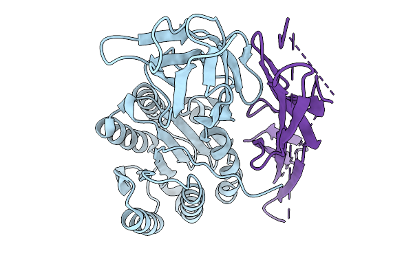 |
Netf - Antxr2 Local Refinement Focused
Organism: Homo sapiens, Clostridium perfringens
Method: ELECTRON MICROSCOPY Release Date: 2026-02-18 Classification: TOXIN |
Organism: Homo sapiens, Clostridium perfringens
Method: ELECTRON MICROSCOPY
Release Date: 2026-02-18
 |
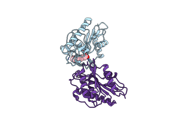
Crystal Structure Of Apt1 Bound To 2-Bromopalmitate
Organism: Homo sapiens
Method: X-RAY DIFFRACTION Resolution:2.10 Å Release Date: 2020-02-05 Classification: HYDROLASE Ligands: J1W |
Organism: Homo sapiens
Method: X-RAY DIFFRACTION
Release Date: 2020-02-05
Ligands: J1W
 |
Crystal Structure Of Apt1 S119A Mutant Bound To Palmitic Acid.
Organism: Homo sapiens
Method: X-RAY DIFFRACTION Resolution:2.60 Å Release Date: 2020-02-05 Classification: HYDROLASE Ligands: PLM |
Organism: Homo sapiens
Method: X-RAY DIFFRACTION
Release Date: 2020-02-05
Ligands: PLM
 |
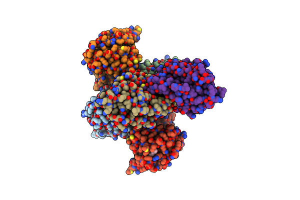
Crystal Structure Of Apt1 C2S Mutant Bound To Palmitic Acid.
Organism: Homo sapiens
Method: X-RAY DIFFRACTION Resolution:2.60 Å Release Date: 2020-02-05 Classification: HYDROLASE Ligands: GOL, PLM |
Organism: Homo sapiens
Method: X-RAY DIFFRACTION
Release Date: 2020-02-05
Ligands: GOL, PLM
 |
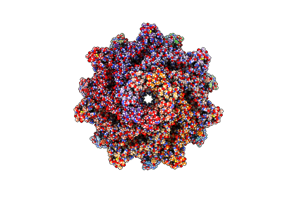
Crystal Structure Of Apt1 Bound To Palmitic Acid.
Organism: Homo sapiens
Method: X-RAY DIFFRACTION Resolution:2.76 Å Release Date: 2020-02-05 Classification: HYDROLASE Ligands: PLM, CL |
Organism: Homo sapiens
Method: X-RAY DIFFRACTION
Release Date: 2020-02-05
Ligands: PLM, CL
 |
Cryo-Em Structure Of Aerolysin Prepore
Organism: Aeromonas hydrophila
Method: ELECTRON MICROSCOPY Release Date: 2016-07-13 Classification: TOXIN |
Organism: Aeromonas hydrophila
Method: ELECTRON MICROSCOPY
Release Date: 2016-07-13
 |
Cryo-Em Structure Of Aerolysin Pore In Lmng Micelle
Organism: Aeromonas hydrophila
Method: ELECTRON MICROSCOPY Release Date: 2016-07-13 Classification: TOXIN |
Organism: Aeromonas hydrophila
Method: ELECTRON MICROSCOPY
Release Date: 2016-07-13
 |
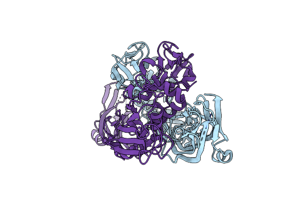
Cryo-Em Structures Of Aerolysin Post-Prepore And Quasipore
Organism: Aeromonas hydrophila
Method: ELECTRON MICROSCOPY Release Date: 2016-07-13 Classification: TOXIN |
Organism: Aeromonas hydrophila
Method: ELECTRON MICROSCOPY
Release Date: 2016-07-13
 |
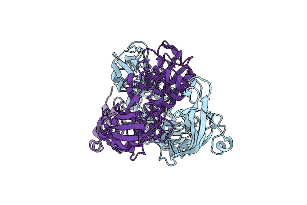
Crystal Structure Of The Activated Aerolysin Mutant H132D
Organism: Aeromonas hydrophila
Method: X-RAY DIFFRACTION Resolution:2.10 Å Release Date: 2010-02-09 Classification: TOXIN |
Organism: Aeromonas hydrophila
Method: X-RAY DIFFRACTION
Release Date: 2010-02-09
 |
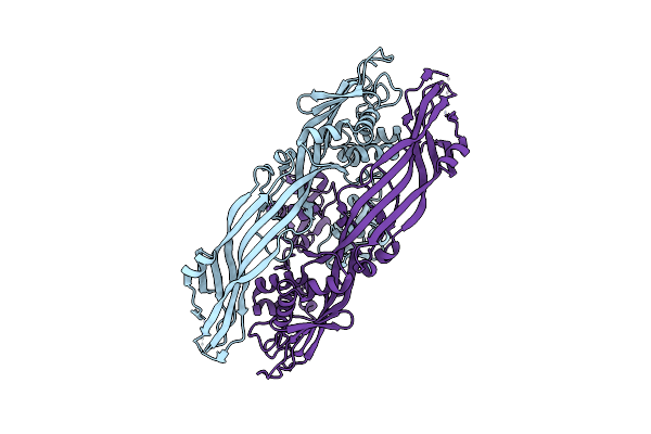
Crystal Structure Of The Activated Aerolysin Mutant H132N
Organism: Aeromonas hydrophila
Method: X-RAY DIFFRACTION Resolution:2.30 Å Release Date: 2010-02-09 Classification: TOXIN |
Organism: Aeromonas hydrophila
Method: X-RAY DIFFRACTION
Release Date: 2010-02-09
 |
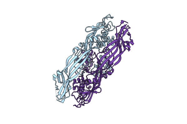
Crystal Structure Of The Proaerolysin Mutant Y221G
Organism: Aeromonas hydrophila
Method: X-RAY DIFFRACTION Resolution:2.88 Å Release Date: 2008-02-12 Classification: TOXIN |
Organism: Aeromonas hydrophila
Method: X-RAY DIFFRACTION
Release Date: 2008-02-12
 |
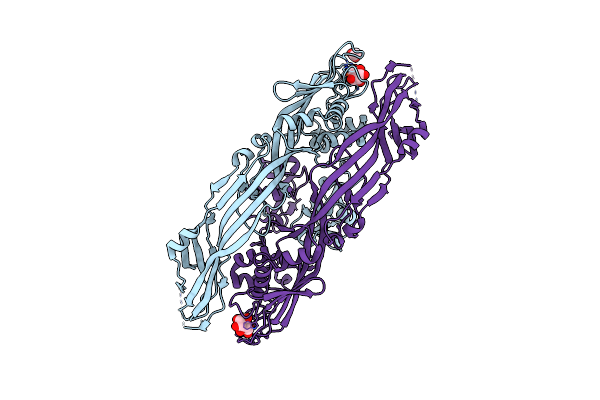
Crystal Structure Of The Proaerolysin Mutant Y221G At 2.2 A
Organism: Aeromonas hydrophila
Method: X-RAY DIFFRACTION Resolution:2.20 Å Release Date: 2008-02-12 Classification: TOXIN |
Organism: Aeromonas hydrophila
Method: X-RAY DIFFRACTION
Release Date: 2008-02-12
 |
Crystal Structure Of The Proaerolysin Mutant Y221G Complexed With Mannose-6-Phosphate
Organism: Aeromonas hydrophila
Method: X-RAY DIFFRACTION Resolution:2.50 Å Release Date: 2008-02-12 Classification: TOXIN Ligands: ACT, M6P |
Organism: Aeromonas hydrophila
Method: X-RAY DIFFRACTION
Release Date: 2008-02-12
Ligands: ACT, M6P