

Search Count: 33
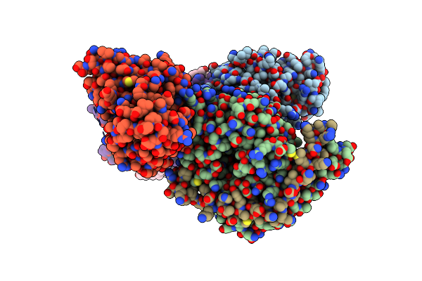 |
Cryo-Em Structure Of Formyl Peptide Receptor 2/Lipoxin A4 Receptor In Complex With Gi
Organism: Homo sapiens, Synthetic construct
Method: ELECTRON MICROSCOPY Release Date: 2020-02-26 Classification: SIGNALING PROTEIN Ligands: CLR, PLM |
Organism: Homo sapiens, Synthetic construct
Method: ELECTRON MICROSCOPY
Release Date: 2020-02-26
Ligands: CLR, PLM
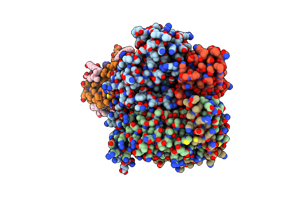 |
Cryo-Em Structure Of Urocortin 1-Bound Corticotropin-Releasing Factor 1 Receptor In Complex With Gs Protein And Nb35
Organism: Homo sapiens, Synthetic construct
Method: ELECTRON MICROSCOPY Release Date: 2020-02-12 Classification: SIGNALING PROTEIN Ligands: CLR, PLM |
Organism: Homo sapiens, Synthetic construct
Method: ELECTRON MICROSCOPY
Release Date: 2020-02-12
Ligands: CLR, PLM
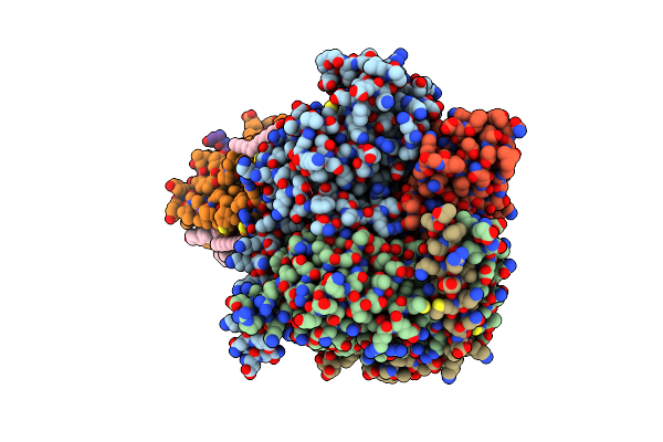 |
Cryo-Em Structure Of Urocortin 1-Bound Corticotropin-Releasing Factor 2 Receptor In Complex With Gs Protein And Nb35
Organism: Homo sapiens, Synthetic construct
Method: ELECTRON MICROSCOPY Release Date: 2020-02-12 Classification: SIGNALING PROTEIN Ligands: CLR, PLM |
Organism: Homo sapiens, Synthetic construct
Method: ELECTRON MICROSCOPY
Release Date: 2020-02-12
Ligands: CLR, PLM
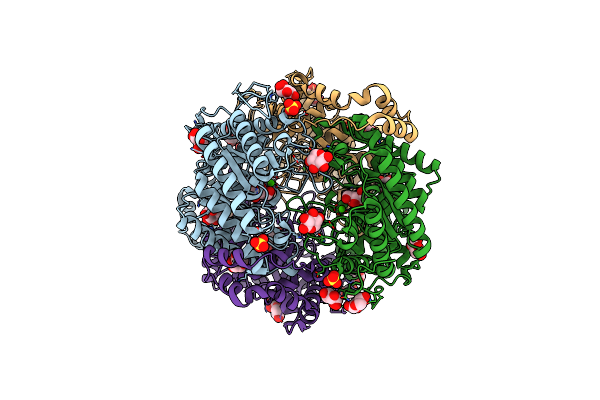 |
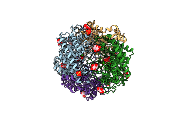
Crystal Structure Of Native Xylose Isomerase From Piromyces E2 Grown In Yeast, In Complex With Xylose
Organism: Piromyces sp. (strain e2)
Method: X-RAY DIFFRACTION Resolution:1.86 Å Release Date: 2020-01-29 Classification: ISOMERASE Ligands: CA, XLS, XYP, XYS, SO4 |
Organism: Piromyces sp. (strain e2)
Method: X-RAY DIFFRACTION
Release Date: 2020-01-29
Ligands: CA, XLS, XYP, XYS, SO4
 |
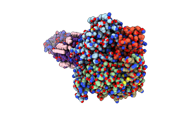
Crystal Structure Of Mutant Xylose Isomerase (V270A/A273G) From Piromyces E2 Grown In Yeast, In Complex With Xylose
Organism: Piromyces sp. (strain e2)
Method: X-RAY DIFFRACTION Resolution:2.00 Å Release Date: 2020-01-29 Classification: ISOMERASE Ligands: CA, XLS, XYP, XYS, SO4 |
Organism: Piromyces sp. (strain e2)
Method: X-RAY DIFFRACTION
Release Date: 2020-01-29
Ligands: CA, XLS, XYP, XYS, SO4
 |
Cryo-Em Structure Of Parathyroid Hormone Receptor Type 1 In Complex With A Long-Acting Parathyroid Hormone Analog And G Protein
Organism: Homo sapiens, Bos taurus, Rattus norvegicus, Synthetic construct
Method: ELECTRON MICROSCOPY Resolution:3.00 Å Release Date: 2019-04-17 Classification: SIGNALING PROTEIN Ligands: CLR, PLM |
Organism: Homo sapiens, Bos taurus, Rattus norvegicus, Synthetic construct
Method: ELECTRON MICROSCOPY
Release Date: 2019-04-17
Ligands: CLR, PLM
 |
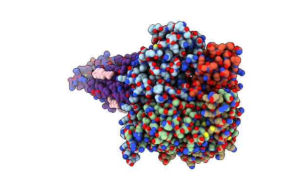
Cryo-Em Structure Of Parathyroid Hormone Receptor Type 1 In Complex With A Long-Acting Parathyroid Hormone Analog And G Protein
Organism: Homo sapiens, Bos taurus, Rattus norvegicus, Synthetic construct
Method: ELECTRON MICROSCOPY Resolution:3.50 Å Release Date: 2019-04-17 Classification: SIGNALING PROTEIN Ligands: CLR, PLM |
Organism: Homo sapiens, Bos taurus, Rattus norvegicus, Synthetic construct
Method: ELECTRON MICROSCOPY
Release Date: 2019-04-17
Ligands: CLR, PLM
 |
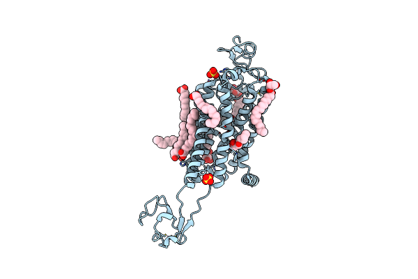
Cryo-Em Structure Of Parathyroid Hormone Receptor Type 1 In Complex With A Long-Acting Parathyroid Hormone Analog And G Protein
Organism: Homo sapiens, Bos taurus, Rattus norvegicus, Synthetic construct
Method: ELECTRON MICROSCOPY Resolution:4.00 Å Release Date: 2019-04-17 Classification: SIGNALING PROTEIN Ligands: CLR |
Organism: Homo sapiens, Bos taurus, Rattus norvegicus, Synthetic construct
Method: ELECTRON MICROSCOPY
Release Date: 2019-04-17
Ligands: CLR
 |
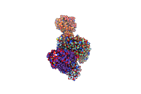
Crystal Structure Of Human Apo-Frizzled4 Receptor
Organism: Homo sapiens, Clostridium pasteurianum
Method: X-RAY DIFFRACTION Resolution:2.40 Å Release Date: 2018-08-22 Classification: MEMBRANE PROTEIN Ligands: ZN, OLC, OLA, SO4, UNX |
Organism: Homo sapiens, Clostridium pasteurianum
Method: X-RAY DIFFRACTION
Release Date: 2018-08-22
Ligands: ZN, OLC, OLA, SO4, UNX
 |
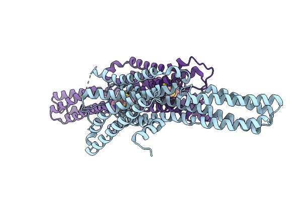
Rhodopsin-Gi Complex
Organism: Escherichia coli, Homo sapiens, Rattus norvegicus, Bos taurus, Synthetic construct
Method: ELECTRON MICROSCOPY Release Date: 2018-06-20 Classification: SIGNALING PROTEIN |
Organism: Escherichia coli, Homo sapiens, Rattus norvegicus, Bos taurus, Synthetic construct
Method: ELECTRON MICROSCOPY
Release Date: 2018-06-20
 |
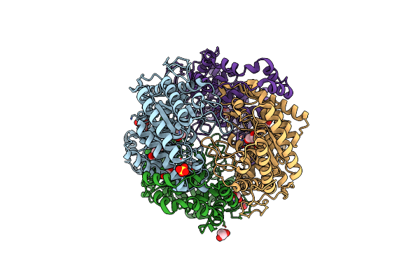
Crystal Structure Of 5-Ht1B Receptor In Complex With Methiothepin
Organism: Homo sapiens, Spodoptera frugiperda
Method: X-RAY DIFFRACTION Resolution:3.90 Å Release Date: 2018-02-07 Classification: ELECTRON TRANSPORT Ligands: 89F |
Organism: Homo sapiens, Spodoptera frugiperda
Method: X-RAY DIFFRACTION
Release Date: 2018-02-07
Ligands: 89F
 |
Crystal Structure Of Xylose Isomerase From Piromyces E2 In Complex With One Mg2+ Ions And Glycerol
Organism: Piromyces sp. e2
Method: X-RAY DIFFRACTION Resolution:1.80 Å Release Date: 2017-11-01 Classification: ISOMERASE Ligands: MG, SO4, GOL |
Organism: Piromyces sp. e2
Method: X-RAY DIFFRACTION
Release Date: 2017-11-01
Ligands: MG, SO4, GOL
 |
Crystal Structure Of Native Xylose Isomerase From Piromyces E2
Organism: Piromyces sp. e2
Method: X-RAY DIFFRACTION Resolution:1.80 Å Release Date: 2017-11-01 Classification: ISOMERASE Ligands: CA, FE2, MG, SO4, GOL |
Organism: Piromyces sp. e2
Method: X-RAY DIFFRACTION
Release Date: 2017-11-01
Ligands: CA, FE2, MG, SO4, GOL
 |
Crystal Structure Of Xylose Isomerase From Piromyces E2 Complexed With One Mg2+ Ion And Xylitol
Organism: Piromyces sp. e2
Method: X-RAY DIFFRACTION Resolution:1.75 Å Release Date: 2017-11-01 Classification: ISOMERASE Ligands: MG, XYL, SO4 |
Organism: Piromyces sp. e2
Method: X-RAY DIFFRACTION
Release Date: 2017-11-01
Ligands: MG, XYL, SO4
 |
Crystal Structure Of Xylose Isomerase From Piromyces E2 In Complex With Two Mg2+ Ions And Xylose
Organism: Piromyces sp. e2
Method: X-RAY DIFFRACTION Resolution:1.90 Å Release Date: 2017-11-01 Classification: ISOMERASE Ligands: MG, XLS, XYP, XYS, SO4 |
Organism: Piromyces sp. e2
Method: X-RAY DIFFRACTION
Release Date: 2017-11-01
Ligands: MG, XLS, XYP, XYS, SO4
 |
Crystal Structure Of Xylose Isomerase From Piromyces E2 In Complex With Two Ca2+ Ions And Xylose
Organism: Piromyces sp. e2
Method: X-RAY DIFFRACTION Resolution:1.86 Å Release Date: 2017-11-01 Classification: ISOMERASE Ligands: CA, XLS, XYP, SO4 |
Organism: Piromyces sp. e2
Method: X-RAY DIFFRACTION
Release Date: 2017-11-01
Ligands: CA, XLS, XYP, SO4
 |
Crystal Structure Of Xylose Isomerase From Piromyces E2 In Complex With Two Mn2+ Ions And Xylose
Organism: Piromyces sp. e2
Method: X-RAY DIFFRACTION Resolution:2.08 Å Release Date: 2017-11-01 Classification: ISOMERASE Ligands: MN, XLS, XYP, SO4 |
Organism: Piromyces sp. e2
Method: X-RAY DIFFRACTION
Release Date: 2017-11-01
Ligands: MN, XLS, XYP, SO4
 |
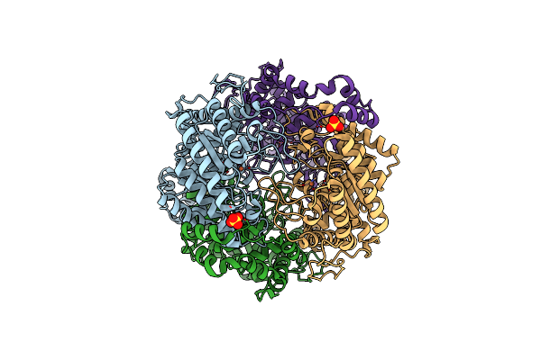
Crystal Structure Of Xylose Isomerase From Piromyces Sp. E2 In Complex With Two Mn2+ Ions And Sorbitol
Organism: Piromyces sp. e2
Method: X-RAY DIFFRACTION Resolution:1.80 Å Release Date: 2017-11-01 Classification: ISOMERASE Ligands: MN, SOR, SO4 |
Organism: Piromyces sp. e2
Method: X-RAY DIFFRACTION
Release Date: 2017-11-01
Ligands: MN, SOR, SO4
 |
Crystal Structure Of Xylose Isomerase From Piromyces E2 In Complex With Two Fe2+ Ions
Organism: Piromyces sp. e2
Method: X-RAY DIFFRACTION Resolution:2.40 Å Release Date: 2017-11-01 Classification: ISOMERASE Ligands: FE2, SO4 |
Organism: Piromyces sp. e2
Method: X-RAY DIFFRACTION
Release Date: 2017-11-01
Ligands: FE2, SO4
 |
Crystal Structure Of Xylose Isomerase From Piromyces E2 In Complex With Two Co2+ Ions And Xylulose
Organism: Piromyces sp. e2
Method: X-RAY DIFFRACTION Resolution:1.93 Å Release Date: 2017-11-01 Classification: ISOMERASE Ligands: CO, XUL, HYP |
Organism: Piromyces sp. e2
Method: X-RAY DIFFRACTION
Release Date: 2017-11-01
Ligands: CO, XUL, HYP