

Search Count: 12
 |
Crystal Structure Of The Gh39 Enzyme From Xanthomonas Axonopodis Pv. Citri
Organism: Xanthomonas axonopodis pv. citri (strain 306)
Method: X-RAY DIFFRACTION Resolution:1.71 Å Release Date: 2020-07-22 Classification: HYDROLASE |
Organism: Xanthomonas axonopodis pv. citri (strain 306)
Method: X-RAY DIFFRACTION
Release Date: 2020-07-22
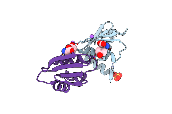 |
Crystal Structure Of Wild-Type Arginine Repressor From The Pathogenic Bacterium Corynebacterium Pseudotuberculosis Bound To Tyrosine
Organism: Corynebacterium pseudotuberculosis (strain c231)
Method: X-RAY DIFFRACTION Resolution:1.69 Å Release Date: 2020-04-22 Classification: DNA BINDING PROTEIN Ligands: TYR, SO4, NA |
Organism: Corynebacterium pseudotuberculosis (strain c231)
Method: X-RAY DIFFRACTION
Release Date: 2020-04-22
Ligands: TYR, SO4, NA
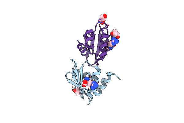 |
Crystal Structure Of Arginine Repressor P115Q Mutant From The Pathogenic Bacterium Corynebacterium Pseudotuberculosis Bound To Arginine
Organism: Corynebacterium pseudotuberculosis (strain c231)
Method: X-RAY DIFFRACTION Resolution:1.70 Å Release Date: 2020-04-22 Classification: DNA BINDING PROTEIN Ligands: ARG, ACT, GOL, TRS |
Organism: Corynebacterium pseudotuberculosis (strain c231)
Method: X-RAY DIFFRACTION
Release Date: 2020-04-22
Ligands: ARG, ACT, GOL, TRS
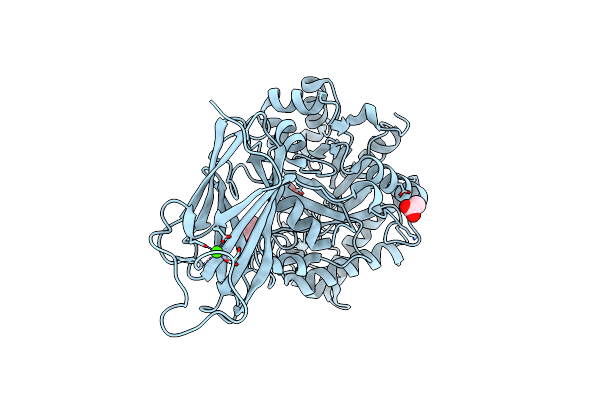 |
Crystal Structure Of A Ruminal Gh26 Endo-Beta-1,4-Mannanase
Organism: Metagenome
Method: X-RAY DIFFRACTION Resolution:1.85 Å Release Date: 2020-03-11 Classification: HYDROLASE Ligands: CA, ACT |
Organism: Metagenome
Method: X-RAY DIFFRACTION
Release Date: 2020-03-11
Ligands: CA, ACT
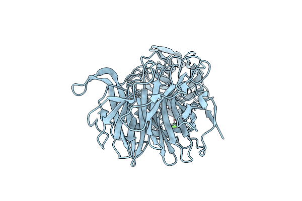 |
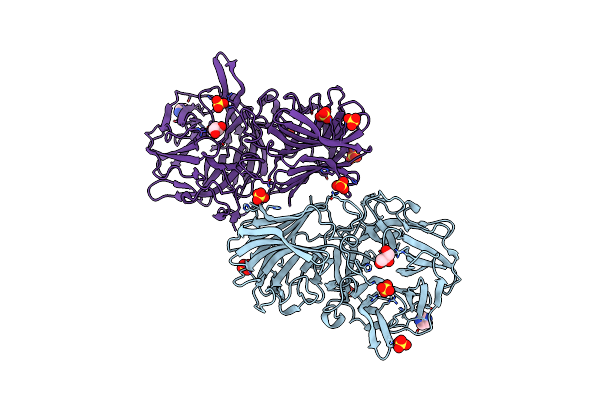
Crystal Structure Of The Gh43 Blxynb Protein From Bacillus Licheniformis
Organism: Bacillus licheniformis (strain atcc 14580 / dsm 13 / jcm 2505 / nbrc 12200 / ncimb 9375 / nrrl nrs-1264 / gibson 46)
Method: X-RAY DIFFRACTION Resolution:2.49 Å Release Date: 2019-04-17 Classification: HYDROLASE Ligands: CA |
 |
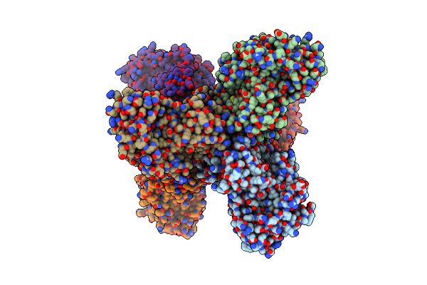
Crystal Structure Of The Gh43 Protein Blxynb Mutant (K247S) From Bacillus Licheniformis
Organism: Bacillus licheniformis (strain atcc 14580 / dsm 13 / jcm 2505 / nbrc 12200 / ncimb 9375 / nrrl nrs-1264 / gibson 46)
Method: X-RAY DIFFRACTION Resolution:1.95 Å Release Date: 2019-04-17 Classification: HYDROLASE Ligands: CA, SO4, GOL, IMD |
 |
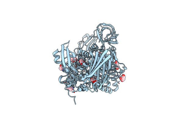
Crystal Structure Of The Gh51 Arabinofuranosidase From Xanthomonas Axonopodis Pv. Citri
Organism: Xanthomonas axonopodis pv. citri (strain 306)
Method: X-RAY DIFFRACTION Resolution:1.91 Å Release Date: 2019-02-20 Classification: HYDROLASE Ligands: GOL |
Organism: Xanthomonas axonopodis pv. citri (strain 306)
Method: X-RAY DIFFRACTION
Release Date: 2019-02-20
Ligands: GOL
 |
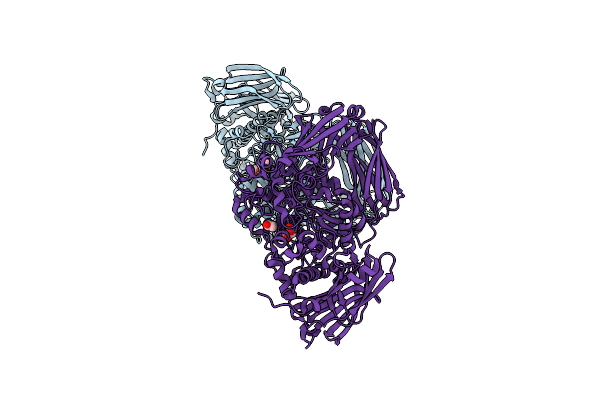
Crystal Structure Of The Gh2 Exo-Beta-Mannanase From Xanthomonas Axonopodis Pv. Citri
Organism: Xanthomonas axonopodis pv. citri (strain 306)
Method: X-RAY DIFFRACTION Resolution:1.90 Å Release Date: 2018-07-18 Classification: HYDROLASE/CARBOHYDRATE Ligands: GOL, PEG, ACT |
Organism: Xanthomonas axonopodis pv. citri (strain 306)
Method: X-RAY DIFFRACTION
Release Date: 2018-07-18
Ligands: GOL, PEG, ACT
 |
Crystal Structure Of The Gh2 Exo-Beta-Mannanase From Xanthomonas Axonopodis Pv. Citri In Complex With Mannose
Organism: Xanthomonas axonopodis pv. citri (strain 306)
Method: X-RAY DIFFRACTION Resolution:2.13 Å Release Date: 2018-07-18 Classification: HYDROLASE/CARBOHYDRATE Ligands: BMA, ACT |
Organism: Xanthomonas axonopodis pv. citri (strain 306)
Method: X-RAY DIFFRACTION
Release Date: 2018-07-18
Ligands: BMA, ACT
 |
Crystal Structure Of The Nucleophile Mutant (E575A) Of The Gh2 Exo-Beta-Mannanase From Xanthomonas Axonopodis Pv. Citri
Organism: Xanthomonas axonopodis pv. citri (strain 306)
Method: X-RAY DIFFRACTION Resolution:2.00 Å Release Date: 2018-07-18 Classification: HYDROLASE/CARBOHYDRATE Ligands: BMA |
Organism: Xanthomonas axonopodis pv. citri (strain 306)
Method: X-RAY DIFFRACTION
Release Date: 2018-07-18
Ligands: BMA
 |
Crystal Structure Of The Acid-Base Mutant (E477A) Of The Gh2 Exo-Beta-Mannanase From Xanthomonas Axonopodis Pv. Citri
Organism: Xanthomonas axonopodis pv. citri (strain 306)
Method: X-RAY DIFFRACTION Resolution:2.20 Å Release Date: 2018-07-18 Classification: HYDROLASE/CARBOHYDRATE Ligands: BMA |
Organism: Xanthomonas axonopodis pv. citri (strain 306)
Method: X-RAY DIFFRACTION
Release Date: 2018-07-18
Ligands: BMA
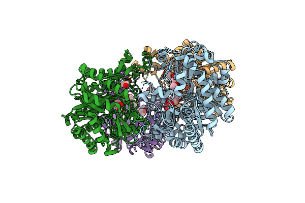 |
Crystal Structure Of A Gh1 Beta-Glucosidase Retrieved From Microbial Metagenome Of Poraque Amazon Lake
Organism: Metagenome
Method: X-RAY DIFFRACTION Resolution:2.75 Å Release Date: 2018-03-07 Classification: HYDROLASE Ligands: GOL, PEG |
Organism: Metagenome
Method: X-RAY DIFFRACTION
Release Date: 2018-03-07
Ligands: GOL, PEG