

Search Count: 31
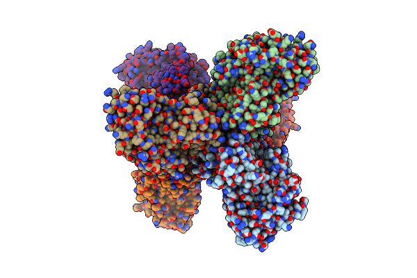 |
Crystal Structure Of The Gh51 Arabinofuranosidase From Xanthomonas Axonopodis Pv. Citri
Organism: Xanthomonas axonopodis pv. citri (strain 306)
Method: X-RAY DIFFRACTION Resolution:1.91 Å Release Date: 2019-02-20 Classification: HYDROLASE Ligands: GOL |
Organism: Xanthomonas axonopodis pv. citri (strain 306)
Method: X-RAY DIFFRACTION
Release Date: 2019-02-20
Ligands: GOL
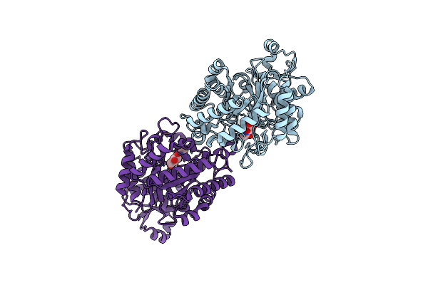 |
Crystal Structure Of The E257A Mutant Of Blman5B In Complex With Glcnac (Co-Crystallization)
Organism: Bifidobacterium longum (strain djo10a)
Method: X-RAY DIFFRACTION Resolution:2.50 Å Release Date: 2019-01-30 Classification: HYDROLASE Ligands: NAG |
Organism: Bifidobacterium longum (strain djo10a)
Method: X-RAY DIFFRACTION
Release Date: 2019-01-30
Ligands: NAG
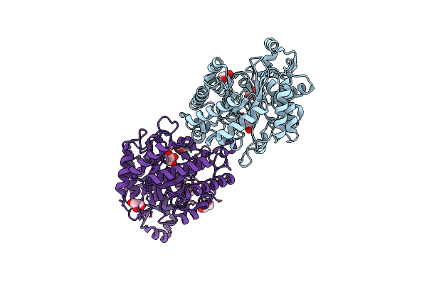 |
Crystal Structure Of Blman5B Solved By Siras
Organism: Bifidobacterium longum djo10a
Method: X-RAY DIFFRACTION Resolution:1.98 Å Release Date: 2019-01-30 Classification: HYDROLASE Ligands: GOL |
Organism: Bifidobacterium longum djo10a
Method: X-RAY DIFFRACTION
Release Date: 2019-01-30
Ligands: GOL
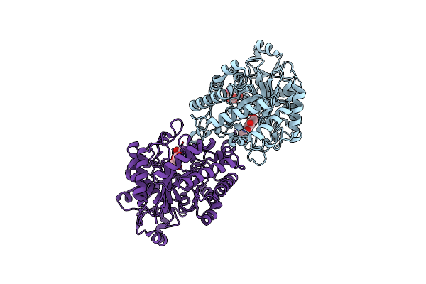 |
Crystal Structure Of The E257A Mutant Of Blman5B In Complex With Glcnac (Soaking)
Organism: Bifidobacterium longum (strain djo10a)
Method: X-RAY DIFFRACTION Resolution:1.75 Å Release Date: 2019-01-30 Classification: HYDROLASE Ligands: NAG, FLC, NA |
Organism: Bifidobacterium longum (strain djo10a)
Method: X-RAY DIFFRACTION
Release Date: 2019-01-30
Ligands: NAG, FLC, NA
 |
Crystal Structure Of Blman5B In Complex With Glcnac (Soaking)
Organism: Bifidobacterium longum (strain djo10a)
Method: X-RAY DIFFRACTION Resolution:1.90 Å Release Date: 2019-01-30 Classification: HYDROLASE Ligands: FLC, NAG |
Organism: Bifidobacterium longum (strain djo10a)
Method: X-RAY DIFFRACTION
Release Date: 2019-01-30
Ligands: FLC, NAG
 |
Crystal Structure Of E257A Mutant Of Blman5B
Organism: Bifidobacterium longum (strain djo10a)
Method: X-RAY DIFFRACTION Resolution:1.80 Å Release Date: 2019-01-30 Classification: HYDROLASE |
Organism: Bifidobacterium longum (strain djo10a)
Method: X-RAY DIFFRACTION
Release Date: 2019-01-30
 |
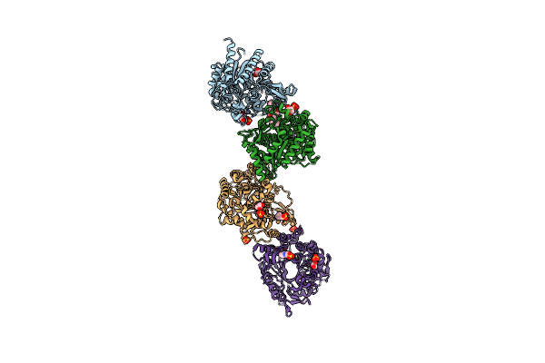
Crystal Structure Of The Gh1 Beta-Glucosidase From Exiguobacterium Antarcticum B7 In Space Group P21
Organism: Exiguobacterium antarcticum (strain b7)
Method: X-RAY DIFFRACTION Resolution:2.24 Å Release Date: 2016-04-13 Classification: HYDROLASE Ligands: SO4 |
Organism: Exiguobacterium antarcticum (strain b7)
Method: X-RAY DIFFRACTION
Release Date: 2016-04-13
Ligands: SO4
 |
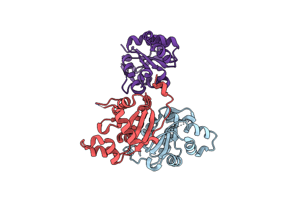
Crystal Structure Of The Gh1 Beta-Glucosidase From Exiguobacterium Antarcticum B7 In Space Group C2221
Organism: Exiguobacterium antarcticum (strain b7)
Method: X-RAY DIFFRACTION Resolution:2.15 Å Release Date: 2016-04-13 Classification: HYDROLASE Ligands: CXS, SO4, GOL |
Organism: Exiguobacterium antarcticum (strain b7)
Method: X-RAY DIFFRACTION
Release Date: 2016-04-13
Ligands: CXS, SO4, GOL
 |
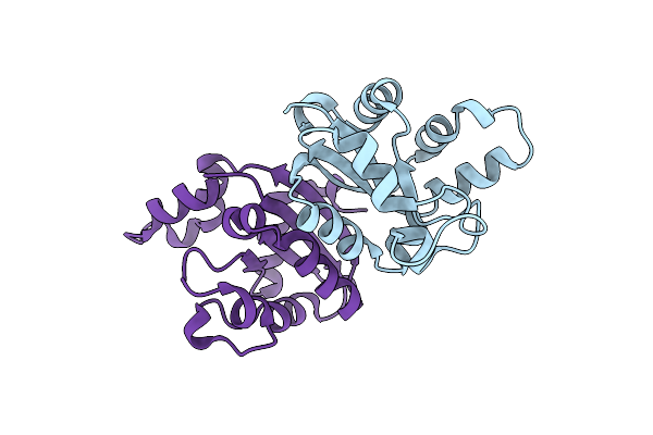
Structure Of Leishmania Nucleoside Diphostate Kinase Mutant P95S
Organism: Leishmania major
Method: X-RAY DIFFRACTION Resolution:3.14 Å Release Date: 2015-10-14 Classification: TRANSFERASE |
Organism: Leishmania major
Method: X-RAY DIFFRACTION
Release Date: 2015-10-14
 |
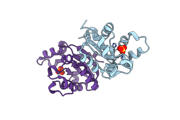
Structure Of Leishmania Nucleoside Diphosphate Kinase Mutant P100S/Del5-Cterm
Organism: Leishmania major
Method: X-RAY DIFFRACTION Resolution:2.30 Å Release Date: 2015-10-14 Classification: TRANSFERASE |
Organism: Leishmania major
Method: X-RAY DIFFRACTION
Release Date: 2015-10-14
 |
Structure Of Leishmania Nucleoside Diphostate Kinase Mutant Del5-Cterm
Organism: Leishmania major
Method: X-RAY DIFFRACTION Resolution:2.95 Å Release Date: 2015-10-14 Classification: TRANSFERASE Ligands: SO4 |
Organism: Leishmania major
Method: X-RAY DIFFRACTION
Release Date: 2015-10-14
Ligands: SO4
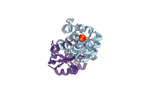 |
Crystal Structure Of The Nucleoside Diphosphate Kinase B From Leishmania Braziliensis
Organism: Leishmania braziliensis
Method: X-RAY DIFFRACTION Resolution:2.70 Å Release Date: 2014-07-09 Classification: TRANSFERASE Ligands: PO4 |
Organism: Leishmania braziliensis
Method: X-RAY DIFFRACTION
Release Date: 2014-07-09
Ligands: PO4
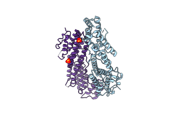 |
Structure Of The Cargo Binding Domain From Human Myosin Va
Organism: Homo sapiens
Method: X-RAY DIFFRACTION Resolution:2.20 Å Release Date: 2013-10-09 Classification: PROTEIN TRANSPORT Ligands: SO4 |
Organism: Homo sapiens
Method: X-RAY DIFFRACTION
Release Date: 2013-10-09
Ligands: SO4
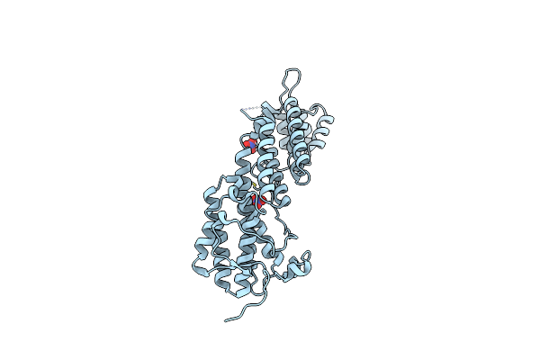 |
Structure Of The Cargo Binding Domain From Human Myosin Vb
Organism: Homo sapiens
Method: X-RAY DIFFRACTION Resolution:2.07 Å Release Date: 2013-10-09 Classification: PROTEIN TRANSPORT Ligands: NO3 |
Organism: Homo sapiens
Method: X-RAY DIFFRACTION
Release Date: 2013-10-09
Ligands: NO3
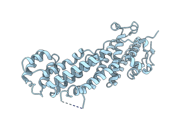 |
Structure Of The Cargo Binding Domain From Human Myosin Vc
Organism: Homo sapiens
Method: X-RAY DIFFRACTION Resolution:2.95 Å Release Date: 2013-10-09 Classification: PROTEIN TRANSPORT |
Organism: Homo sapiens
Method: X-RAY DIFFRACTION
Release Date: 2013-10-09
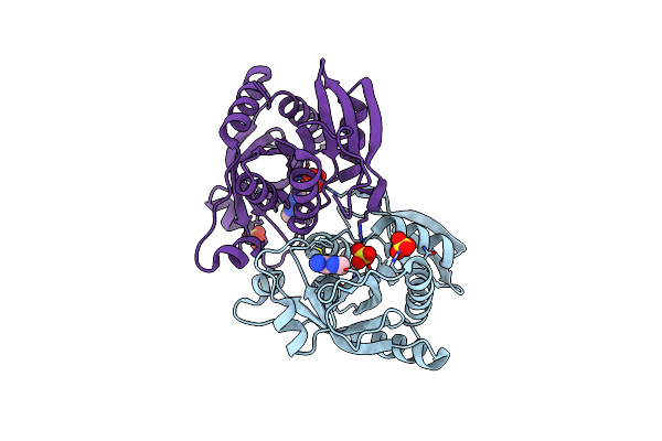 |
Crystal Structure Of The Hexameric Purine Nucleoside Phosphorylase From Bacillus Subtilis At Ph 4.2
Organism: Bacillus subtilis
Method: X-RAY DIFFRACTION Resolution:2.35 Å Release Date: 2012-09-26 Classification: TRANSFERASE Ligands: SO4, ADE |
Organism: Bacillus subtilis
Method: X-RAY DIFFRACTION
Release Date: 2012-09-26
Ligands: SO4, ADE
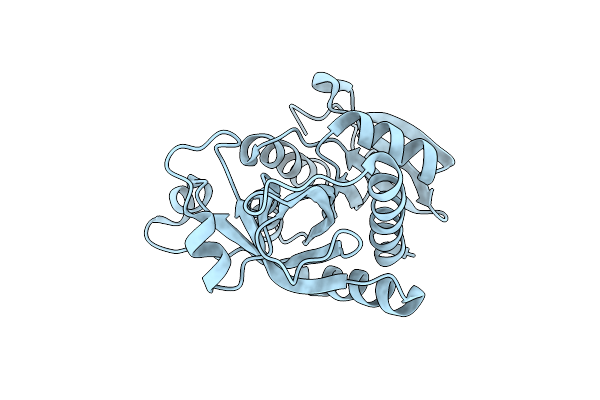 |
Crystal Structure Of The Hexameric Purine Nucleoside Phosphorylase From Bacillus Subtilis In Space Group P6322 At Ph 4.6
Organism: Bacillus subtilis
Method: X-RAY DIFFRACTION Resolution:2.65 Å Release Date: 2012-09-26 Classification: TRANSFERASE |
Organism: Bacillus subtilis
Method: X-RAY DIFFRACTION
Release Date: 2012-09-26
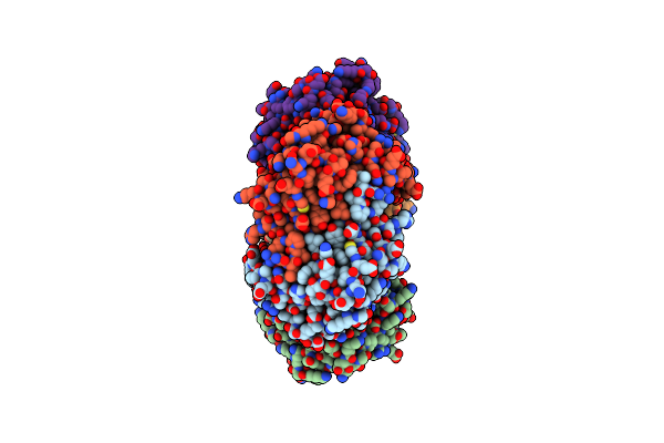 |
Crystal Structure Of The Hexameric Purine Nucleoside Phosphorylase From Bacillus Subtilis In Space Group P212121 At Ph 5.6
Organism: Bacillus subtilis
Method: X-RAY DIFFRACTION Resolution:1.61 Å Release Date: 2012-09-26 Classification: TRANSFERASE Ligands: SO4, GOL |
Organism: Bacillus subtilis
Method: X-RAY DIFFRACTION
Release Date: 2012-09-26
Ligands: SO4, GOL
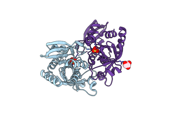 |
Crystal Structure Of The Hexameric Purine Nucleoside Phosphorylase From Bacillus Subtilis In Space Group H32 At Ph 7.5
Organism: Bacillus subtilis
Method: X-RAY DIFFRACTION Resolution:1.70 Å Release Date: 2012-09-26 Classification: TRANSFERASE Ligands: SO4, CL, GOL |
Organism: Bacillus subtilis
Method: X-RAY DIFFRACTION
Release Date: 2012-09-26
Ligands: SO4, CL, GOL
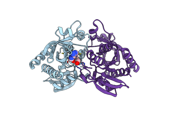 |
Crystal Structure Of The Hexameric Purine Nucleoside Phosphorylase From Bacillus Subtilis In Complex With Adenosine
Organism: Bacillus subtilis
Method: X-RAY DIFFRACTION Resolution:1.91 Å Release Date: 2012-09-26 Classification: TRANSFERASE Ligands: ADN |
Organism: Bacillus subtilis
Method: X-RAY DIFFRACTION
Release Date: 2012-09-26
Ligands: ADN